What is the oral cavity?
The oral cavity is the 1st part of the digestive tube. It extends anteroposteriorly from the lips to the oropharyngeal isthmus. The oral cavity is used for the ingestion of food and fluids.
Parts of oral cavity
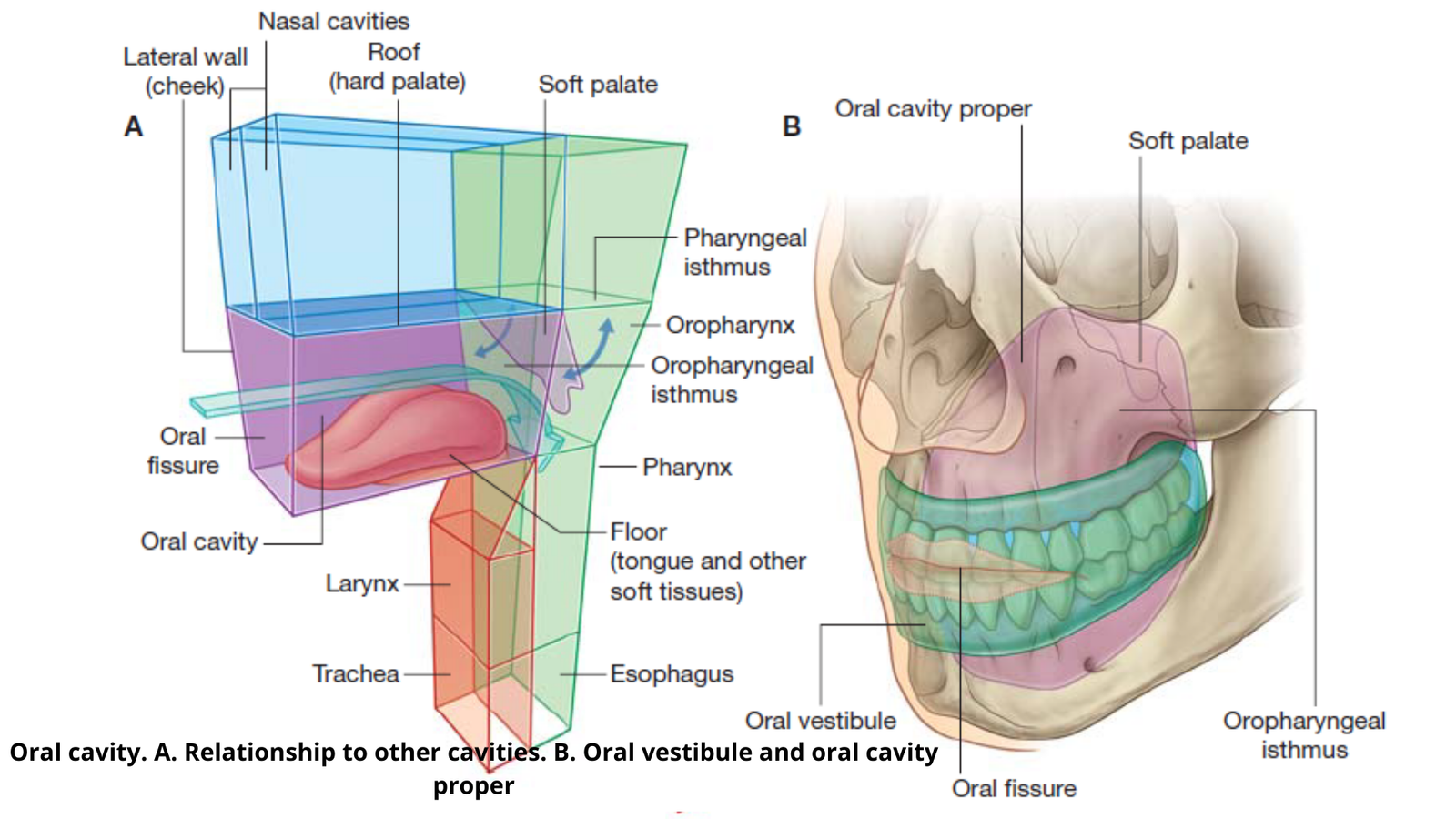
The oral cavity is divided into two parts:
- Vestibule (slit-like space between lips/cheeks and teeth/gingivae(gums).
- The oral cavity proper: larger space inside the teeth and gums.
Vestibule of the mouth
The vestibule is a narrow space that lies outside the teeth and gums, and inside the lips and cheeks. It is limited above and below by the reflection of the mucus membrane from the lips and cheeks to the gums.
When the mouth is open, it freely communicates with the oral cavity proper. When it is closed i.e. when teeth are occluded, it communicates on each side with the oral cavity proper through a small gap called the retromolar region (behind the third molar teeth and ramus of the mandible).
Except for teeth, the entire vestibule line by mucus membrane. Anteriorly and laterally bounded by lips and cheek.
The cheeks made up of buccinator muscle. Opening in the vestibule of the mouth;
- Opening of the parotid duct.
- Opening of labial and buccal mucus glands.
- Opening of 4 or 5 molar glands (mucus) situated on the buccopharyngeal fascia.

Lips
Lips are anterior two mobile musculofibrous folds. Upper and lower lips meet laterally at an angle of the mouth, usually present in front of the first premolar tooth.
Lips are internally lined by mucus membrane and externally lined by skin. The mucocutaneous junction lines the edge of the lip. The red portion of the lip is called the vermillion zone.
Vermillion border: skin and vermillion zone meet. Frenulum of the lip: an internal aspect of each lip is connected to the corresponding gum by a median fold of the mucus membrane.
Structure of lips
Superficial to deep:
- Skin.
- Superficial fascia.
- Orbicularis Oris muscle.
- Submucosa containing mucus glands.
- Mucous membrane.
The blood supply in the lips:
- Arterial supply: labial branches of the facial artery.
- Venous drainage: labial branches of the facial vein.
- Lymphatic drainage:
- Submental lymph node (central part of the lower lip).
- Submandibular lymph node (lateral part of the lower and upper lip in mouth area).
- Nerve supply:
- Sensory supply: trigeminal nerve.
- Upper lip: The labial branches of the infraorbital nerve which is a branch of maxillary division.
- Lower lip: mental nerve (branch of mandibular division)
- The red portion of the lips is highly sensitive.
Cheeks
The cheeks are fleshy flaps forming a huge part of the face. Each cheek is continuous in front of the lip. Nasolabial sulcus or the nasolabial furrow: junction between the two.
It extends from the side of the nose to the angle of the mouth. Cheeks are largely composed of buccinator muscle is covered by buccopharyngeal fascia. It contains buccal glands, blood vessels, and nerves.
A layer of the cheek (From superficial to deep):
- Skin.
- Superficial fascia containing some muscles of facial expression, viz. zygomaticus major, risorius.
- Buccal pad of fat.
- Buccopharyngeal fascia.
- Buccinator muscle between the alveolar processes of both jaws.
- Submucosa containing buccal (mucus) glands.
- Mucous membrane.
Gums/gingiva
It is fibrous tissue covered with a smooth vascular mucous membrane. The envelop (cover) the alveolar processes of the jaws & the necks of the teeth.
At the necks of the teeth, the fibrous tissue of the gum begins to be continuous with the periodontal membrane, which attaches the teeth to their sockets.
Parts of gum:
- Free part (free gingiva): Surrounds the neck of the tooth like a collar.
- Attached part (attached gingiva): Firmly attached to the alveolar process.
- Interdental part (interdental gingiva): Extension of the attached gingiva between the teeth.
Nerve supply:
- Upper gums on:
- The labial aspect is supplied by the posterior, middle, and anterior superior alveolar nerves. •lingual aspects are supplied by the greater palatine and nasopalatine nerves.
- Lower gums on:
- Labial aspects are supplied by the buccal branch of the mandibular nerve, and an incisive branch of the mental nerve.
- Lingual aspects are supplied by the lingual nerves.
Lymphatic Drainage:
- Upper gums: submandibular lymph nodes.
- Lower gums: submental lymph nodes and submandibular lymph nodes.
Applied anatomy
- Gingivitis
- Scurvy
Oral cavity proper
The roof and a floor present in the oral cavity proper. Posteriorly the oral cavity communicates with the oropharynx through the oropharyngeal isthmus (also called isthmus of fauces). Boundaries of Oral cavity proper:
- Superiorly: soft palate.
- Inferiorly: tongue.
- Each side: palatoglossal arches
The floor of the mouth
A small horseshoe-shaped region. It is situated beneath the anterior two-thirds of the tongue and above the muscular diaphragm formed by two mylohyoid muscles.
The surface of the floor is formed by a mucus membrane, which connects the tongue to the mandible. Laterally the mucus membrane travels from the side of the tongue onto the mandible.
Anteriorly the mucus membrane stretches from one half of the mandible to the other. The anterior part of the floor is called the sublingual region, which intervenes between the ventral surface of the anterior two-thirds of the tongue and the floor of the mouth.
Sublingual region
The lesser surface of the tongue is attached or connected to the floor of the mouth by a median fold of the mucus membrane known as frenulum linguae.
On each side of the lower end of the frenulum, there is an elevation called sublingual papilla, on the summit of which opens the submandibular duct.
The sublingual gland projects up into the floor of the mouth and produces an elevation in the mucus membrane on each side of the frenulum called the sublingual fold. The majority of the sublingual ducts open on this fold.
The roof of the mouth
It is formed by the palate. Anterior two-thirds of the palate made up of bones is called the hard palate. The posterior 1/3rd part made of the soft tissue is called the soft palate.
In the posterior-free margin of the soft palate, a small conical projection called uvula hangs down in the median region.
A poorly marked median raphe extends from the uvula to the incisive papilla -a slight elevation behind the incisive fossa.
The mucus membrane in the anterior part of the hard plate is thrown into 3 or 4 transverse palatine folds but posteriorly it is comparatively smooth.
[embeddoc url=”https://notesmed.com/wp-content/uploads/2021/01/Oral-cavity.pdf” download=”all”]