What is sternocleidomastoid muscle?
The sternocleidomastoid muscle is the principal muscle of the neck region. It obliquely extends across the side of the neck region & dividing the anterior and posterior triangle of the neck.
Origin and insertion
Origin:
- Sternal head: Upper part of the anterior surface of the manubrium of the sternum.
- The clavicular head of SCM: Superior surface of medial 1/3rd of the clavicle.
Insertion
- Sternal head: Lateral one-half of superior nuchal line.
- The clavicular head of SCM: lateral surface of the mastoid process.

Nerve supply
- Accessory nerve [ XI ]
- Branches from anterior rami of C2 to C3 (C4).
Blood supply
Arterial supply
- Upper part: Posterior auricular artery & occipital artery.
- Middle part: Superior thyroid artery.
- Lower part: Suprascapular artery.
Actions of the sternocleidomastoid muscle
- Unilateral contraction
- Bilateral contraction
Individually — will tilt head toward the shoulder area on the same side rotating head to turn face to the opposite side; acting together, draw head forward.
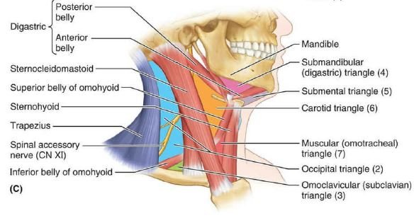
Relations of the sternocleidomastoid muscle
- The investing layer of deep cervical fascia present outside the sternocleidomastoid muscle.
- Four sternomastoid arteries and spinal accessory nerve pierced this muscle.
- There are the following superficial & deep relations are below;
Superficial Relations
- Skin
- Platysma
- Three cutaneous nerves
- Great auricular
- Transverse cervical
- Medial supraclavicular
- Lesser occipital
- External jugular vein
- Superficial cervical lymph nodes
- Parotid gland
Deep Relations
In the upper part
- Muscle:
- Posterior belly of digastric
- Longissimus capitis
- Splenius capitis
- Artery: Occipital artery
In the middle part
- Muscles:
- Levator scapulae muscle
- Scalenus anterior muscle
- Scalenus medius muscle
- Scalenus posterior muscle
- Splenius capitis muscle
- Inferior belly of the omohyoid muscle
Arteries:
- Common carotid artery
- Internal carotid artery
Veins:
- Internal jugular vein
- anterior jugular vein
Nerves:
- Vagus nerve
- Spinal accessory nerve
- Cervical plexus nerve
- Brachial plexus (upper part)
- Ansa cervicalis (inferior root)
Glands:
- The thyroid gland
- Lymph nodes
In the lower part
Muscles:
- Sternohyoid muscle
- Sternothyroid muscle
- Scalenus anterior muscle
Arteries:
- Suprascapular artery
- Transverse cervical artery
Veins:
- Anterior jugular veins
Nerves:
- Brachial plexus (lower part)
- Phrenic nerve
Applied anatomy
- Torticollis or wry neck
- Sternomastoid tumor
Torticollis or wry neck
- It occurs due to a spasm of the trapezius muscle and sternocleidomastoid muscle in the neck.
- Spasmodic torticollis:
- Repeated painful contraction muscles
- Caused by exposure to cold & maladjustment of pillow during sleep.
- Reflex torticollis:
- It occurs due to irritation of the spinal accessory nerve caused by inflamed or suppurating lymph nodes.
- Congenital torticollis:
- Due to birth injury to muscles.
Sternomastoid tumor
- The Middle Third of the sternocleidomastoid muscle swelling due to edema & ischemic necrosis caused by trauma at birth.