What is a scalp?
The scalp is the soft tissue covering the cranial vault of the skull and the name of the scalp is derived from the layer of the scalp in the skull.
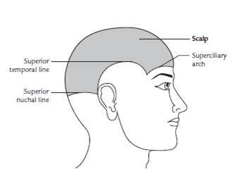
Extent of scalp
- Anterior: Supraorbital margins (up to the eyebrows)
- Posterior: external occipital protuberance and superior nuchal lines
- On each side: zygomatic arch (up to superior temporal line)
Layers of the scalp
The scalp consists of the following five layers (SCALP) from superficial to deep, they are;
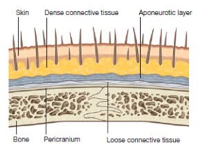
- Skin
- Connective tissue (superficial fascia)
- Aponeurosis {deep fascia in the form of occipitofrontalis & its aponeurosis-epicranial aponeurosis(galea aponeurotica)}
- Loose areolar tissue
- Pericranium
Skin
The skin on the outermost layer of the scalp is thick and hairy except over the forehead. They are firmly attached to the epicranial aponeurosis through the dense connective tissue of the superficial fascia.
Being a hairy area it contains a maximum number of hairy follicles & an associated large number of sebaceous glands and also contains numerous sweat glands, and so the commonest site of sebaceous cysts.
Connective tissue (superficial fascia)
The superficial fascia of the scalp is made of dense connective(fibrofatty) tissue. It binds the skin to subjacent aponeurosis. It provides a medium for the passage of blood vessels and nerves.
Wounds bleed profusely as blood vessels are prevented or stopped from retraction by fibrous tissue. Bleeding is stopped by applying pressure against the underlying bone. The inflammation causes little swelling but is much more painful.
Epicranial aponeurosis
This layer is made by occipitofrontalis muscle and its aponeurosis (galea aponeurosis). Anteriorly frontal belly and posteriorly occipital belly of occipitofrontalis muscle are inserted. The frontal belly originates from the skin of the forehead and mingled with the orbicularis oculi muscle. Occipital belly originates from lateral 2/3rd of superior nuchal line.
Loose areolar tissue
It is made of loose areolar tissue and serves as a natural plane of cleavage during craniotomy. It extends anteriorly into the eyelids because the frontalis has no bony attachment and posteriorly to the superior nuchal line.
And on each side – attachment of the aponeurosis to the temporal fascia. The bleeding causes generalized swelling of the scalp of the skull and bleeding leads to a black eye. It is a dangerous layer of the scalp due to emissary veins open here and carries any infections inside the brain (intracranial dural venous sinuses).
Pericranium
It is the fifth layer of the scalp of the vault of the skull and formed by the periosteum of bones vault of the skull known as pericranium. It is loosely attached to the surface of the bone but is firmly adherent to the sutures, which in turn attaches it to the endocranium. The injury deep to it takes the shape of the bone called cephalhematoma.
Blood supply of the scalp
Arterial supply
- Supratrochlear artery
- Supraorbital artery
- Superficial temporal artery
- Posterior auricular artery
- Occipital artery
Venous drainage
- Supratrochlear veins
- Supraorbital veins
- Superficial temporal veins
- Posterior auricular veins
- Occipital veins
Lymphatics drainage
- Anterior part: Preauricular (parotid) group of lymph node
- Posterior part: Posterior (mastoid) group of lymph node occipital group of lymph node
Nerve supply of the scalp
- In front of the auricle
- Supratrochlear nerve
- Supraorbital nerve
- Zygomaticotemporal nerve
- Auriculotemporal nerve
- The temporal branch of the facial nerve
- Behind auricle
- Greater auricular nerve
- Lesser occipital nerve
- Greater occipital nerve
- Third occipital nerve
- Posterior Auricular branch of the facial nerve
Applied anatomy
- Black eye
- Safety-valve hematoma
- Cephalhematoma
- Caput succedaneum
- Scalp laceration