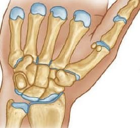
What is the wrist joint?
The wrist joint is a synovial joint of ellipsoid variety between the lower end of the radius bone and carpus bone.
Articular surfaces wrist joint
- Proximal articular surface: the inferior surface of the lower extremity of the radius bone and inferior surface of the triangular articular disc of inferior radio-ulnar joint
- Distal articular surface: proximal surfaces of the scaphoid bone, lunate bone, and triquetral bones.
Ligaments of the wrist joint
- Capsular ligaments (joint capsule)
- Radial collateral ligament
- Ulnar collateral ligament
- Palmar radio-carpal ligament
- Palmar ulnocarpal ligament
- Dorsal radio-carpal ligament
Capsular ligament (joint capsule)
The fibrous covering of the wrist joint in your hand and is attached above to the distal ends of the radius bone and ulna bone, and below to the proximal row of carpal bones. The synovial membrane lines the inner surface area of the fibrous capsule and extends or elongates up to the margins of the articular surfaces.
Radial collateral ligament
It extends or elongates from the tip of the styloid process of radius bone to lateral aspects of the scaphoid and trapezium bone. It is also related to the radial artery in the hand region.
Ulnar collateral ligament
It elongates or extends from the tip of the styloid process of the ulnar bone to the medial aspects of the triquetral and pisiform bones.
Palmar radio-carpal ligament
It extends from the anterior margin of the lower extremity (end) of the radius bone to the anterior surfaces of the scaphoid, lunate bone, and triquetral bones. It is formed due to the condensing (thickening) of the lateral part of the anterior aspect of the fibrous capsule.
Dorsal radio-carpal ligament
It extends downwards and medially from the posterior margin of the lower extremity of the radius bone to the dorsal surface of the scaphoid, lunate bone, and triquetral bones.
Palmar ulnocarpal ligament
It elongates vertically downwards from the base of the styloid process & adjoining part of the articular disc to the anterior surface of the lunate bone and triquetral bones. It is formed due to the thickening or condensing of the medial part of the anterior aspect of the fibrous capsule of the joint.
Relations in the wrist joint
Anterior:
- Tendons of flexor digitorum superficialis muscle, flexor digitorum profundus muscle, and related synovial sheath (ulnar bursa).
- The tendon of flexor pollicis longus muscle and related synovial sheath (radial bursa).
- Median nerve.
- Tendon of flexor carpi radialis muscle & associated synovial bursa in the joint.
- Ulnar nerve.
Posterior:
- Extensor tendons of wrist area or joint and fingers, and associated synovial sheaths.
- The anterior interosseous artery.
- The anterior interosseous nerve.
Lateral:
- Radial artery (across the radial collateral ligament in the hand).
- Tendon of abductor pollicis longus (APL) muscle.
- Tendon of extensor pollicis brevis (EPB) muscle.
Medial:
- Dorsal cutaneous branch of the ulnar nerve in the joint.
Applied anatomy
- Ganglion (Gk = swelling or knot): It is a non-tender cystic swelling or bulging, which sometimes commonly on the dorsal aspect of the wrist joint in your hand.
[embeddoc url=”https://notesmed.com/wp-content/uploads/2020/08/ELBOW-JOINT.pdf” download=”all” cache=”off”]