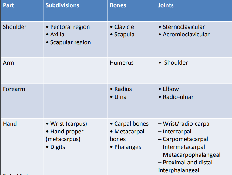
Parts of the upper limb:
[embeddoc url=”https://notesmed.com/wp-content/uploads/2020/08/The-Upper-limb.pdf” download=”all”]
The upper limb can be divided into the following four parts, they are: 1)Shoulder 2) Arm (brachium) 3)Forearm or ante-brachium 4)Hand.
Shoulder Region:
The shoulder region includes: (a)Axilla or armpit, (b)Scapular region or parts around the scapula (shoulder blade), and (c)Pectoral or breast region on the front of the chest.
The bones of the shoulder region are (a) the Clavicle (collar bone) and (b)The scapula (shoulder blade).
These bones are articulate with each other at the acromioclavicular joint and form the shoulder girdle. The shoulder girdle articulates with the rest of the skeleton of the body only at the small sternoclavicular joint.

Arm or brachium:
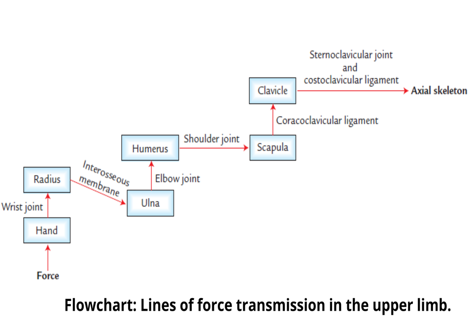
It is a part of the upper limb between the shoulder and elbow (or cubitus). The bone of the arm is the humerus, which articulates with the scapula at the shoulder joint and upper ends of radius and ulna bones at the elbow joint.
Forearm or Ante-brachium:
It is a part of the upper limb between the elbow and the wrist. The bones of the forearm are radius and ulna. These bones articulate with the lower end of the humerus at the elbow joint and with each other forming radioulnar joints.
Hand/Manus:
The hand/manus consists of the following parts of the body, they are: (a)wrist or carpus, (b)Hand proper (or metacarpus), and (c)Digits (thumb and fingers).
The wrist:
The wrist (carpus) consists of eight carpal bones arranged in two rows, and each row consisting of four bones.
@ She Looks Too Pretty. Try To Catch Her.
Scaphoid, lunate, triquetral, pisiform, trapezium, trapezoid, capitate, and hamate.
The carpal bones articulate;
- with each other to form intercarpal joints
- proximally with radius forming radiocarpal/ wrist joint, and
- distally with metacarpal bones to form carpometacarpal joints
Had proper:
It consists of five metacarpal bones numbered one to five from lateral to medial side in anatomical position. They articulate;
- proximally with distal row of carpal bones forming carpometacarpal joints
- with each other forming intermetacarpal joints, and
- distally with proximal phalanges forming metacarpophalangeal joints.
Digits:
The digits are five in number and numbered 1 to 5 from lateral to the medial side. The first digit is called the thumb and the remaining four digits are called fingers.
Each digit is supported by three short long bones (the phalanges) except the thumb, which is supported by only two phalanges.
The phalanges form metacarpophalangeal joints with metacarpals and interphalangeal joints with one another.
The first carpometacarpal joint has a separate joint cavity hence movements of the thumb are much free than that of any digit/finger.
Muscle of the upper limb:
The muscles include:
(a)the muscles that attach the limb and girdle to the body and
(b)the muscles of the arm, forearm, and hand.
The deltoid muscle covers the shoulder like a hood and is commonly used for intramuscular injections.
The arm and forearm are invested in the deep fascia like a sleeve and are divided into anterior and posterior compartments by intermuscular septa.
The muscles of anterior and posterior compartments mainly act synergistically to carry out specific functions. The muscles of the anterior compartment are mainly flexors and those of posterior compartment extensors.
The muscles of the hand are responsible for their various skilled movements such as grasping, etc.
Nerve supply:
The nerve supply to the upper limb is derived from the brachial plexus. The brachial plexus is formed by ventral rami of C5 to C8 and T1 spinal nerves. The five main branches of the brachial plexus, are;
- Axillary nerve: Supplies the deltoid and Teres minor muscle.
- Musculocutaneous nerve: Anterior (flexor) compartments of the arm and forearm.
- Median nerve: Anterior (flexor) compartments of the arm and forearm.
- Ulnar nerve: Anterior (flexor) compartments of the arm and forearm.
- Radial nerve: Posterior compartments (extensor) of the arm and forearm.
N.B. All the intrinsic muscles of the hand are supplied by the ulnar nerve except muscles of the thenar eminence and the first two lumbrical muscles.
Arteries of the limb:
It is supplied by four main arteries:
- The axillary artery supplies the shoulder region.
- The brachial artery supplies the anterior and posterior compartments of the arm.
- The radial and ulnar arteries: It supplies the lateral and medial parts of the forearm, respectively.
Venous drainage:
The deep veins of the upper limb follow the arteries and run superiorly towards the axilla, where the axillary vein travels superiorly and becomes the subclavian vein at the outer border of the 1st rib.
The subclavian vein continues towards the root of the neck where it joins the internal jugular vein to form the brachiocephalic vein.
The two brachiocephalic veins (right and left) join each other to form superior vena cava, which drains into the heart.
The superficial veins of the upper limb originate from the dorsal venous arch of the hand.
The lateral end of the dorsal venous arch forms the cephalic vein, which runs along the lateral aspect of the upper limb and terminates into the axillary vein in the axilla.
The medial end of the dorsal venous arch forms the basilic vein, which ascends along the medial aspect of the upper limb and empties into the axillary vein as well.
Anterior to the elbow, the cephalic vein is connected to the basilic vein via the median cubital vein.
Lymphatic drainage:
The lymphatics of the upper limb originating in the hand. The superficial lymph vessels follow the superficial veins. The deep lymph vessels follow the deep arteries (viz. radial, ulnar, and brachial) and pass superiorly to the axilla where they drain into the axillary lymph nodes.
Applied anatomy:
Dislocation of the shoulder joint (most commonly dislocated joint in the body), elbow joint, and lunate bone of the hand.
Fractures of the clavicle (most commonly fractured bone in the body), humerus, radius, and scaphoid.
The scaphoid is the most commonly fractured bone of the hand.
Nerve injuries of brachial plexus, median nerve, radial nerve, and ulnar nerve.