Thorax
The thorax is the upper part of the trunk of the body. It extends from the root of the neck region to the abdomen. The chest is utilized as a synonym for the thorax. The cavity of the trunk is divided by the diaphragm into an upper part which is called the thoracic cavity and the lower part is called the abdominal cavity.
The thoracic cavity contains the principal or primary organs of respiration called the lungs, which are separated from each other by bulky & movable median septum which is called the mediastinum. The principal structures present in the mediastinum are the heart and great vessels in the human.
Thoracic cage
The thorax is supported by a skeletal framework which is called the thoracic cage. The thoracic cage provides attachment to the various muscles of the thorax, upper extremities, back region, and diaphragm. It is osteocartilaginous (bone and cartilage) and elastic in nature.
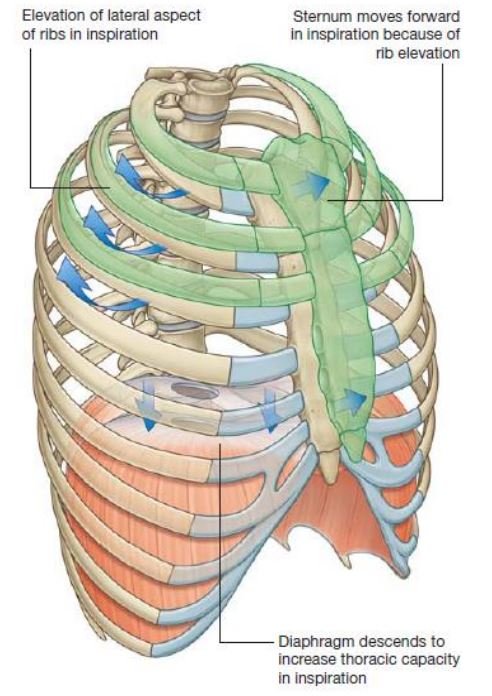
It is principally designed for increasing or decreasing the intrathoracic pressure in the thorax so that air is sucked into the lungs during inspiration & expelled out from the lungs during the expiration process. It is essential for the mechanism of the respiration process.

Formation of the thoracic cage
- Anteriorly: Sternum (breast bone).
- Posteriorly: Thoracic (T1 to T12) vertebrae and intervening intervertebral discs present on it.
- Laterally each side: 12 pairs of ribs involved and associated 12 pairs of costal cartilages in the body.
- The rib cage is formed by the sternum, costal cartilages, and ribs which are attached to the thoracic vertebrae.
The ribs articulate as following:
- Posteriorly: All the ribs articulate with the thoracic vertebrae in the human.
- Anteriorly:
- The upper seven ribs (1st to 7th) articulate with the side of the sternum throughout their costal cartilages.
- 8th, 9th, and 10th articulate with each other throughout their costal cartilages.
- 11th and 12th do not articulate with sternum and anterior ends of their costal cartilages are free.
The shape of the thoracic cage:
It bears a resemblance to a truncated cone with its narrow extremity (end) above and broad extremity (end) below. The narrow superior end is continuous above with the root of the neck from which it is partly separated or divided on either side by the suprapleural membranes.
The broad inferior (lower) end is completely separated or divided from the abdominal cavity by the diaphragm but provides passage to structures like the aorta, esophagus, and inferior vena cava(IVC). The diaphragm is a dome-shaped structure with its convexity directed upwards. The upper abdominal viscera located within the thoracic cage and is protected by it.
Transverse section of the thorax
| The thoracic cavity in adult | A thoracic cavity in infant |
| Kidney shaped | Circular |
| Ribs obliquely placed | Ribs horizontally placed |
| Transverse diameter can be increased by thoracic breathing process (Hence respiration is thoracoabdominal) | Transverse diameter cannot be increased by thoracic breathing process(Hence respiration is purely abdominal) |
Superior thoracic aperture (Thoracic inlet)-Thorax
The thoracic cavity communicates with the root of the neck through a narrow opening which is called a superior thoracic aperture or thoracic inlet due to the fact that air and food enter the thorax through the trachea and esophagus, respectively.
Boundaries of Superior thoracic aperture (Thoracic inlet):
- Anteriorly: Superior (upper) border of manubrium sterni.
- Posteriorly: Anterior border of the superior surface of the body of the first thoracic (T1) vertebra.
- Laterally (on each side): Medial border of the first rib with its cartilage.
The superior (upper) end of the anterior boundary lies 1.5 inches below the superior (upper) end of the posterior boundary due to the fact that the first rib slopes downwards and forwards from its posterior end to the anterior end. The superior (upper) border of the manubrium sterni which is lies at the level of the upper border of the third thoracic (T3) vertebra.
Shape and Dimensions:
- Shape: Reniform/kidney-shaped.
- Dimensions:
- Transverse diameter: 4.5 inches.
- Anteroposterior diameter: 2.5 inches.
The diaphragm of the superior thoracic aperture/(suprapleural membrane or Sibson’s fascia):
The part of the thoracic inlet, on either side, is closed with a thick fascial sheet which is known as suprapleural membrane or Sibson’s fascia, or diaphragm of the superior thoracic aperture which is a tent-shaped structure.,
Attachments:
- The apex of Sibson’s fascia: Tip of the transverse process of the seventh (C7) vertebra.
- The base of Sibson’s fascia: Inner border of the first rib with its costal cartilage.
Relations:
- Superior surface: The subclavian vessels.
- Inferior surface: Cervical pleura, covering the apex of your lungs.
Functions of Sibson’s fascia:
- It preserves the underlying cervical pleura, below which lies the apex of the lung.
- It resists the intrathoracic pressure during the respiration period. As a result, the root of the neck is not puffed (out of the breath) up and down during the respiration period.
N.B. Morphologically, Sibson’s fascia represents the spread (layout) out degenerated tendon of the scalenus minimus (or pleuralis) muscle.
Structures Passing Through Thoracic Inlet
Muscles:
- Sternohyoid.
- Sternothyroid.
- Longus cervicis/longus colli.
Arteries:
- Right and left internal thoracic arteries.
- Brachiocephalic trunk/artery.
- Left common carotid artery.
- Left subclavian artery.
- Right and left superior intercostal arteries.
Nerves:
- Right and left vagus nerves.
- Left recurrent laryngeal nerve.
- Right and left phrenic nerves.
- Right and left first thoracic nerves.
- Right and left sympathetic chains.
Veins:
- Right and left brachiocephalic veins.
- Right and left 1st posterior intercostal veins.
- Inferior thyroid veins.
Lymphatics: –Thoracic duct.
Others structures:
- Anterior longitudinal ligament.
- Esophagus.
- Trachea.
- Right and left domes of cervical pleura.
- Apices of right and left lungs.
Inferior thoracic aperture(Thoracic outlet)
It is wide and surrounds the upper (superior) part of the abdominal cavity. The large musculoaponeurotic diaphragm which is attached to the margins of the thoracic outlet separates or differentiates the thoracic cavity from the abdominal cavity.
Boundaries:
- Anteriorly: Xiphisternal joint.
- Posteriorly: Body of 12th thoracic vertebra.
- Laterally (on each side): Costal margin & 11th and 12th ribs.
The diaphragm of inferior thoracic aperture:
The thoracic outlet is closed by a huge dome-shaped musculotendinous which is called the diaphragm. Since it separates or differentiates the thoracic cavity from the abdominal cavity, it is also known as the thoracoabdominal diaphragm.
The diaphragm is the principal muscular organ of respiration. It is a dome-shaped structure and consists of a peripheral muscular part and a central fibrous part which is called a central tendon.
Applied anatomy:
[embeddoc url=”https://notesmed.com/wp-content/uploads/2020/08/Thorax.pdf” download pdf file=”all” cache=”off”]