What is the pterygopalatine fossa?
The pterygopalatine fossa is an inverted (upturn) teardrop-shaped pyramidal gap (space) & presents deeply under the apex of the orbit, between the pterygoid process of sphenoid bone beyond and the perpendicular plate of palatine in front.
And more literally, the back of the maxilla replaces the palatine bone as the anterior boundary of the entrance of the fossa which is known as the pterygomaxillary fissure.

Boundaries of Pterygopalatine fossa
The walls of the pterygopalatine fossa are formed by the following parts of the palatine bone, maxilla, and sphenoid bones.
- Anterior:
- Perpendicular plate of the palatine bone-in anteriorly
- The posterior surface of the maxillary bone (superomedial part).
- Posterior:
- Pterygoid process
- Adjoining part of the anterior surface of the greater wing bone of the sphenoid.
- Medial:
- The superior part of the perpendicular plate of the palatine orbital bone
- Sphenoidal process of the palatine bone medially.
- Lateral:
- The fossa opens into the infratemporal fossa throughout the pterygomaxillary fissure laterally.
- Superior:
- Beneath the surface of the body of the sphenoid bone and orbital process of the palatine bone.
- The lateral part of the roof is open and here fossa is opened into the orbit through an inferior orbital fissure.
- Inferior:
- The pyramidal process of the palatine bone in the angle in the middle of the maxilla and the pterygoid process.
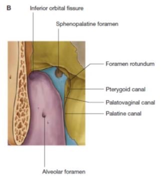
Communications
Anteriorly: It communicates with the orbit via. the medial end of the inferior orbital fissure.
Posteriorly:
1. It communicates with the middle cranial fossa via. the foramen rotundum.
2. It communicates with the foramen lacerum via. the pterygoid canal.
3. It communicates with the pharynx via. the palatovaginal canal.
Medially: With the nose via. the sphenopalatine foramen.
Laterally: With the infratemporal fossa via. the pterygomaxillary fissure.
Inferiorly: With the oral cavity via. greater and lesser palatine canals.
Contents
The main contents of the pterygopalatine fossa in the following structures:
- Maxillary nerve.
- Pterygopalatine ganglion.
- The 3rd part of the maxillary artery
Golden points:
1) The principal muscle of the infratemporal fossa is the Lateral pterygoid muscle.
2) All the key muscles of mastication close the mouth except the Lateral pterygoid muscle, which helps to open the mouth.
3) Clinically the most principal branch of the maxillary artery is the middle meningeal artery.
4) Peripheral heart in the area of the head and neck is the Lateral pterygoid muscle.
5) The principal elevator of the lower jaw is the Masseter muscle.
6) The most usual nerve block given in dentistry is the Inferior alveolar nerve block.
7) The most usual dislocation of the temporomandibular joint is Anterior dislocation.