What is a popliteal fossa?
The popliteal fossa is a principal area of transition present between the thigh and leg and is the major route by which structures pass from one region to the other region.
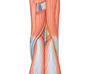
The popliteal fossa is a diamond-shaped hollow present on the back of the knee joint & becomes prominent when the knee is flexed. It is an important anatomical area due to the fact that it provides passage for the main vessels and nerves from the thigh to the leg.
Boundaries of a popliteal fossa
The popliteal fossa is bounded by the following structures given below:
- Superomedially: Semitendinosus muscle & semimembranosus muscle
- Superolaterally: Biceps femoris muscle
- Inferomedially: Medial head of the gastrocnemius muscle
- Inferolaterally: The lateral head of the gastrocnemius muscle is supplemented by the plantaris muscle.
- Floor (or anterior wall):
It is formed from above downward by
- The popliteal surface of the femur.
- Knee joint capsule & oblique popliteal ligament.
- The popliteal fascia covering the popliteus muscle.
- Roof (or posterior wall):
It is formed from the strong popliteal fascia and the superficial fascia over the roof that contains the following structures;
- Short saphenous vein.
- Three cutaneous nerves:
- The terminal part of the posterior cutaneous nerve of the thigh region
- The posterior division of the medial cutaneous nerve of the thigh region
- Sural communicating nerve
The roof is penetrated by all these structures except the posterior division of the medial cutaneous nerve of the thigh.
Contents of the popliteal fossa
- Popliteal artery and its branches
- Popliteal vein and its tributaries
- Tibial nerve and its branches
- Common peroneal nerve and its branches
- Popliteal lymph nodes
- Popliteal pad of fat
- The posterior cutaneous nerve of the thigh in the terminal part
- Descending the genicular branch of the obturator nerve in this region
- The terminal part of the short saphenous vein
Clinical correlation
Popliteal pulse:
To feel the popliteal pulse in this region, at first flex the knee joint to relax the popliteal fascia & then place the fingertips of both hands in the popliteal fossa with thumbs resting on the patient’s patella.
Popliteal pulse is the toughest (difficult) pulse to feel amongst all the peripheral pulses.
Popliteal aneurysm:
The popliteal artery is more prone to an aneurysm than any other artery in the body. Clinically, the popliteal aneurysm mostly presents as a pulsatile midline swelling in the popliteal fossa.
Baker’s cyst:
It is cystic swelling that occurs in the popliteal fossa because of inflammation of synovial bursa situated directly below the tendon of semimembranosus or protrusion of synovial membrane of the cavity of the knee joint throughout the fibrous capsule of the knee joint.
[embeddoc url=”https://notesmed.com/wp-content/uploads/2020/08/Popliteal-fossa.pdf” download=”all”]