
What is mediastinum?
The mediastinum is a middle space present in the thoracic cavity between the two pleural sacs of the body.
The region present between the two pleural sacs bounded anteriorly by the sternum, posteriorly by the thoracic vertebrae, superiorly by the thoracic inlet, and inferiorly by the diaphragm. It is subdivided into the superior and inferior mediastinum.
Boundaries
- Anteriorly: sternum
- Posteriorly: vertebral column (12 thoracic vertebrae)
- Superiorly: thoracic inlet
- Inferiorly: diaphragm
- On each side: mediastinal pleura.
Division
Trans-thoracic plane: An imaginary plane line passing from the sternal angle anteriorly to the inferior border of the body of the T4 vertebra that divides the mediastinum into the superior and inferior mediastinum.
Superior mediastinum
Boundaries
- Infront : Manubrium Sterni.
- Behind: upper 4 thoracics vertebrae with intervertebral discs in the body.
- Above: Thoracic inlet present
- Below: Trans-thoracic plane which is passing through the sternal angle anteriorly to the lower border of the 4th thoracic vertebra posteriorly.
Contents of the superior division
- Muscles: sternohyoid, sternothyroid, and longus colli
- Thymus gland
- Veins: right and left brachiocephalic vein, superior vena cava (SVC), and left superior intercostal vein.
- Arteries: Arch of aorta brachiocephalic artery, left common carotid, and left subclavian vein.
- Nerve: Vagus, Phrenic, left recurrent laryngeal nerve.
- Trachea and esophagus
- Thoracic duct
- Different groups of lymph nodes; paratracheal, brachiocephalic, and tracheobronchial lymph nodes.
Inferior Mediastinum
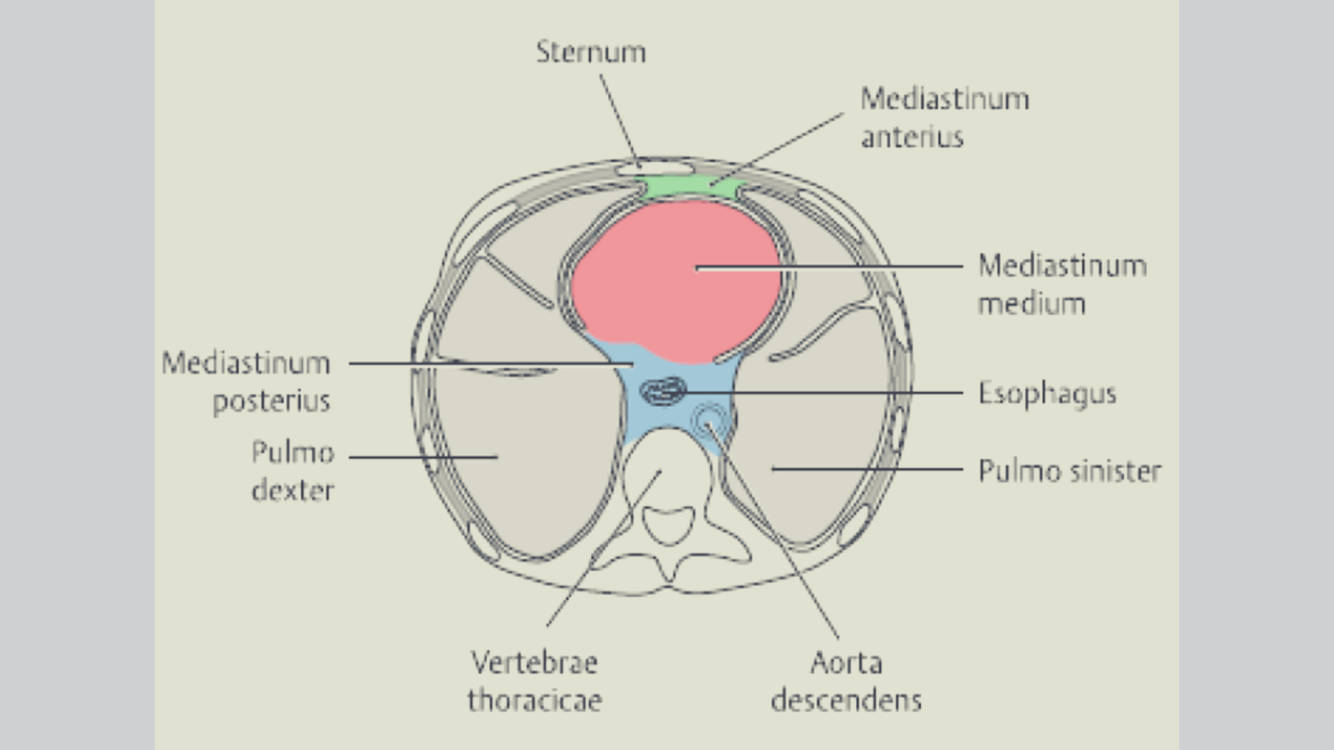
The pericardium further divides the inferior mediastinum into the anterior which is in front of the pericardium. and posterior is behind the pericardium. The middle mediastinum is the pericardium and its contents.
Anterior Mediastinum
It is a narrow space present, overlapped by the anterior border of both lungs.
Boundaries
- Anteriorly: a body of sternum
- Posteriorly: pericardium
- Superiorly: imaginary plane.
- Inferiorly: the superior surface of the diaphragm.
- On each side: mediastinal pleura.
Contents
- Sternopericardial ligaments
- Lymph nodes with lymphatics
- Small mediastinal branches of the internal thoracic artery
- The lowest part of the thymus
- Areolar tissue
Middle Mediastinum
It is the widest subdivision of the inferior mediastinum and occupied by the pericardium and its contents. It is limited space on each side by the mediastinal pleura.
Contents
- Heart and pericardium
- Ascending aorta and pulmonary trunk and pulmonary arteries.
- The lower part of the superior vena cava.
- Four pulmonary veins.
- Arch of the azygos vein.
- Phrenic nerve
- Pericardiophrenic vessels.
- The terminal part of the trachea, and bronchi.
- Deep cardiac plexus of nerves
- Inferior tracheobronchial lymph nodes.
Posterior Mediastinum
Boundaries
- Anteriorly: pericardium, bifurcation of the trachea, pulmonary vessels, and posterior part of the upper surface of the diaphragm.
- Posteriorly: 8 thoracic vertebrae and intervertebral disc.
- On each side: mediastinal pleura.
- Above: imaginary horizontal plane
- Below: the diaphragm
- On each side : mediastinal pleura.
Contents
- Oesophagus
- Descending aorta
- Azygos vein and Hemi azygos vein
- Thoracic duct
- Vagus nerve
- Splanchnic nerves
- Posterior mediastinal lymph nodes.
Applied anatomy
- Deflection of mediastinum
- Mediastinitis—a spread of the infection from the neck most commonly.
- Mediastinal tumor and the cysts.
- Mediastinoscopy.
- Mediastinal syndrome.
[embeddoc url=”https://notesmed.com/wp-content/uploads/2020/10/MEDIASTINUM.pdf” download=”all”]

Thank you for this informative post! I found the detailed breakdown of the mediastinum subdivisions very helpful, especially the emphasis on clinical relevance in applied anatomy. It really enhanced my understanding of its importance in thoracic anatomy. Looking forward to more posts like this!