Overview of the trigeminal nerve
The trigeminal nerve is the largest cranial nerve and contains both sensory and motor fibers in the human. It is sensory to the greater part of the head region & motor to mainly the muscles of mastication.

The functional component of the trigeminal nerve
- General somatic afferent (GSA)
- GSA carries general sensations from the skin of the face and scalp area, teeth, gum, oral cavity & nasal cavities with paranasal sinuses, cornea, conjunctiva, and most of the dura mater in human.
- The cell bodies of neurons for general sensations are situated in the trigeminal ganglion.
- Carry proprioceptive sensation from the mastication muscles & extraocular muscles of the eye.
- The cell bodies are situated in the mesencephalic nucleus of the 5th cranial nerve.
- Special visceral efferent (SVE)
- Conveyed by the motor root to provides the muscles which are developed from the 1st branchial arch including muscles of mastication, tensor tympani & tensor velli palatini, mylohyoid and anterior belly of the digastric muscle.
Nuclei of the trigeminal nerve
- Sensory nucleus (main)
- Spinal nucleus
- Mesencephalic nucleus
- Motor nucleus
Main sensory nucleus
It located in the posterior part of the pons and continuously beneath the spinal nucleus and it carries fibers from trigeminal ganglion by sensory roots (sensation of touch and pressure).
Spinal nucleus
It spread (extends) from the caudal end of the main sensory nucleus to the 2nd or 3rd cervical segments of the spinal cord. It receives the pain and temperature sensation from all trigeminal areas via the sensory root.
Mesencephalic nucleus:
It presents a lateral part of the grey matter throughout the cerebral aqueduct. It contains cell bodies of the unipolar first sensory neurons for the proprioceptive impulses from the mastication muscles, face, and extraocular muscle of the human eye.
The sensory component of the trigeminal nerve
- Sensations of pain, temperature, touch, and pressure from the skin area of the face and mucous membranes in the body travel throughout the axons whose cell bodies are located in the semilunar or trigeminal sensory ganglion.
- Central processes of these cells form the huge sensory root of the trigeminal nerve in the body.
- About half the fibers separate into ascending and descending branches when they enter the pons in the body.
- The remainder ascends or descends without division in the nerve.
- The ascending branches terminate(wind up) in the major sensory nucleus, and the descending branches terminate in the spinal nucleus.
- Proprioceptive impulses from the masticatory muscles & from the facial muscles and extraocular muscles are carried by fibers in the sensory root of the trigeminal nerve that have bypassed into the semilunar or trigeminal ganglion.
Motor nucleus and its communication
- In the pons medial to the main sensory nucleus in the body.
- Receives corticonuclear fibers from both cerebral hemispheres in the brain of the human being.
- And it also receives from the reticular formation, tectum, red nucleus, and medial longitudinal fasciculus in the body.
- Supplies the masticatory muscles, tensor tympani, tensor veli palatini, mylohyoid, and anterior belly of the digastric muscle in the body.
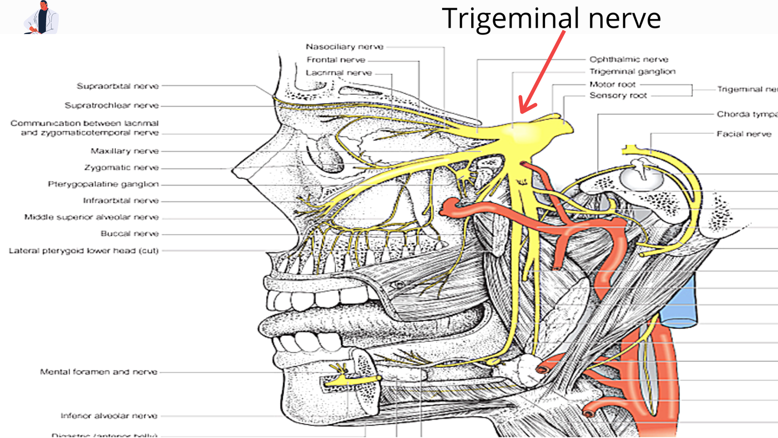
A course of the trigeminal nerve
It gives up the anterior side of the pons & then passes forwarding out of the posterior cranial fossa. It rests on the upper (superior) surface area of the apex of the petrous part of the temporal bone in the middle cranial fossa.
The sensory root enlarges to form the crescent-shaped trigeminal ganglion- lies (located) within a pouch of dura mater- trigeminal or Meckel’s cave in the body.
Division of trigeminal nerve
There are three divisions of the nerve that arise from the anterior border of the ganglion in the body.
- Ophthalmic division– leaves the skull throughout the superior orbital fissure.
- Maxillary division– foramen rotundum.
- Mandibular division– foramen ovale.
Ophthalmic division:- tip and side of the nose area, upper eyelid, and forehead in the body.
Maxillary division:- upper lip, side and ala of the nose area, lower eyelid, the upper part of the cheek, and a small portion of the temple in the body.
Mandibular division– lower lip, chin, skin area overlying the mandible except for its angle, cheek, part of the pinna and external acoustic meatus, and most of the temple.

Applied anatomy
Trigeminal neuralgia (tic douloureux):
It is a clinical case that presents as paroxysmal episodes of severe pain of sudden onset & short duration in the area of distribution of one or more of the three divisions of the trigeminal nerve in the body.
In these clinical conditions, the ophthalmic division (CN V1) is not commonly involved. The most commonly trigeminal neuralgia is associated with maxillary (CN V2) and mandibular divisions (CN V3) of the trigeminal nerve in the body. It is often associated with dental caries in the human.
- Sensory function tested by using a small part of cotton and a pin over each area of the face. In the area of the lesion of ophthalmic division cornea and conjunctiva will be insensitive to touch.
- Motor function tested s done by asking the patient to clench the teeth.
[embeddoc url=”https://notesmed.com/wp-content/uploads/2020/10/trigeminal-nerve.pdf” download=”all”]