The submandibular gland is a walnut-size irregular in shape gland. It is a mixed gland that secretes both mucus as well as serous but predominantly serous secretion in the body.
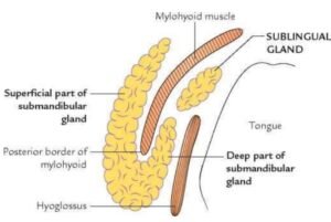
The weight of the submandibular gland is around 10 to 20 grams. It consists of two parts a larger superficial and a smaller deep part, and it continues with each other around the posterior border of the mylohyoid muscle.
Position: It located partially in the digastric triangle & partially in the submandibular fossa up to the mylohyoid line.
Parts
Superficial part: It is a large part that lies superficial to the mylohyoid muscle.
Deep part: It is a small part that lies deep in the mylohyoid muscle.
These two parts of the gland are continuous with each other around the region of the posterior border of the mylohyoid muscle.
Superficial part
Parts
Surfaces: Includes inferior surface, lateral surface, and medial surfaces.
Two ends: Includes anterior end and posterior end.
Anterior end: It extends or elongates up to the anterior belly of the digastric muscle.
Posterior end: It extends or elongates up to the stylomandibular ligament, which separates the submandibular gland from the parotid gland in the body.
This extremity presents a groove produced by the ascending limb of the cervical loop of the facial artery.
Relations:Superficial surface (inferior surface): from superficial to deep is surrounded by the following structures listed below;
Skin.
Superficial fascia that containing platysma and a cervical branch of the facial nerve in the body.
Deep fascia.
Facial vein.
Submandibular lymph nodes.
Lateral surface:
Submandibular fossa present on the inner aspect of the body of the mandible.
Medial pterygoid muscle (near its insertion).
Facial artery grooves in the posterosuperior part then come out from the lower border of the mandible.
Medial surface: Extensive and divided into the following three parts.
(a) Anterior part is related to:
Mylohyoid muscle.
Submental branch of the facial artery.
Mylohyoid nerve and its vessels.
(b) Middle (intermediate) part is related to:
Hyoglossus muscle.
Styloglossus muscle.
Lingual and hypoglossal nerves.
Submandibular ganglion.
(c) Posterior part is related to:
Styloglossus muscle.
Stylohyoid ligament.
Glossopharyngeal nerve.
Wall of the pharynx.
Deep part of the submandibular gland
It is small and extends or elongates forward in the interval between the mylohyoid muscle & hyoglossus up to the posterior end of the sublingual salivary gland.
Relations:
Laterally – mylohyoid muscle.
Medially – hyoglossus muscle.
Above – lingual nerve & submandibular ganglion.
Below – the hypoglossal nerve goes along with a pair of veins (vena comitans nervi hypoglossi).
Submandibular duct (Wharton’s duct)
It is about 5 cm long duct in human beings. The duct begins from several tributaries in the superficial part of the submandibular gland and comes out from the medial surface of this part of the gland beyond the posterior border of the mylohyoid muscle.
It traverses the deep part of the gland, between mylohyoid muscles and hyoglossus muscles. It next passes between the sublingual gland and genioglossus muscle to open in the floor of the mouth on the top of the sublingual papilla at the side of the frenulum of the tongue in the human body.
Blood supply: by branches of the facial arteries & lingual arteries with corresponding veins.
The lymph vessels drainages into submandibular nodes and then the jugulo-omohyoid node in the body.
Nerve supply
Parasympathetic (secretomotor) supply:
The preganglionic parasympathetic nerve fibers which are arisen from the superior salivatory nucleus (SSN) in the pons and they pass successively through the facial nerve, chorda tympanic nerve, and lingual nerves; and that terminates in the submandibular ganglion, which gives out a relay station.
The postganglionic nerve fibers arise from this ganglion and they directly supply the submandibular gland.
Sympathetic supply:
The preganglionic nerve fibers which are arisen from the T1 spinal segment and enter the cervical sympathetic trunk to pass on its superior cervical sympathetic ganglion.
The postganglionic nerve fibers which are arisen from the superior cervical sympathetic ganglion, form plexus throughout the facial artery and thus reach the gland through this artery.
Sensory supply: lingual nerve.
Applied aspect
The submandibular gland may be enlarged due to obstruction or inflammation of its duct by calculus or tumors.
The gland is palpated by bimanual examination- when a finger in the floor of the mouth & another finger below the angle of the mandible.