Define the spinal cord?
The spinal cord is the lower elongated cylindrical part of the central nervous system (CNS) in the human body. The length is about 45cm and the weight is around 30 grams.
The location of the spinal cord is in the upper 2/3rd part of the vertebral canal in the human body. It extent at the level of the foramen magnum to the lower border of the first lumbar (L1) vertebrae.
- Conus medullaris: The spinal cord tapers rapidly toward the inferior to form a cone-shaped termination.
- Filum terminale: The filament of connective tissue that descending from the apex of conus medularis.
- Filum terminale internum: Superior 15 cm which reaches up to the inferior border of S2 vertebrae in the human being.
- Filum terminale externum: Inferior 5 cm which fuses with dura mater and reaches up to the first coccyx vertebra in the body of a human being.
Coverings of spinal cord
Spinal meninges i.e.
- Dura mater
- Arachnoid &
- Pia mater
Modification of pia mater to form the following structure
- Linea splendens
- Ligamentum denticulatum
- Filum terminale.
Positional changes of spinal cord
- Up to 3rd month of intrauterine life: Spinal cord extends throughout the length of the vertebral column in the body. The spinal nerve lies at the identical level as the corresponding intervertebral foramen of the body.
- At birth: Spinal cord up to the inferior border of the third lumbar (L3) vertebrae.
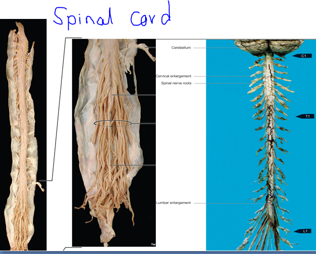
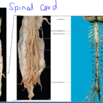
External features of spinal cord
- Fissures and sulci
- Attachment of spinal nerves
- Enlargements
- Cauda equina.
Fissures and sulci
The surface of the cord is divided into two symmetrical halves by the anterior median fissure (anteriorly) & posterior median sulcus (posteriorly).
Each half of the cord is subdivided into the following parts such as anterior, lateral, and posterior areas by an anterolateral and postero-lateral sulcus.
Attachments of spinal nerve
There are 31 pairs of spinal nerves that come out from the side of the spinal cord of the body. The spinal nerves are mixed nerves and each nerve consisting of ventral and dorsal root & Spinal ganglion and also Ventral and dorsal rami present.
- Cervical -8
- Thoracic – 12
- Lumbar -5
- Sacral- 5
- Coccygeal- 1
- Spinal segment
A portion of the spinal cord that furnishes the dorsal and ventral rootlet to a single pair of a spinal nerve in the body. The spinal segments do not correspond with the vertebral segment of the body.
- Cervical region-add 1
- Up to T6-add 2
- T7-T10- add 3
- T11-L3 spinal segment.
- T12- S1 spinal segment.
- L1- overlies the rest of the lumbosacral segment.
Cauda equina
Nerve roots of coccygeal nerves, sacral nerves, & lumbar nerves from the caudal part of the cord that takes more or less a vertical course and forms a bunch of nerve fibers around the filum terminale are known as cauda equina.
Its resemblance to the tail of a horse so-called cauda equina. The cauda equina consists of the roots of the lower four pairs of lumbar nerves, five pairs of sacral nerves, and one pair of coccygeal nerves.
Enlargements
- Cervical enlargement:
- C5-T1.
- To accommodate more motor neurons to supply muscles of the arm in the body
- Lumbo-sacral enlargement: L2-S3.
[embeddoc url=”https://notesmed.com/wp-content/uploads/2020/10/Spinal-cord.pdf” download=”all”]