- Radiation therapy: Radiologists use high-power X-rays to help decrease the risk of the tumour coming back again when surgery is undergone. Again and again, people have radiation therapy before surgery, so that the surgeon can remove less amount of tissue. But in the case of RMS radiation therapy is typically given along with chemotherapy.
- Chemotherapy: Anti-cancer drugs kill cancer cells all around the body. Most chemotherapy is delivered injected into a vein or taken by mouth (orally). In some clinical cases, doctors use chemotherapy before surgery because it makes the tumour smaller. Doctors or consultants may recommend chemotherapy to kill any cancer cells left behind after surgery.
- For the low-risk group people, the main combinations of drugs used are:
- VA: Actinomycin-D (vincristine and dactinomycin)
- VAC: vincristine, dactinomycin, and cyclophosphamide
- For the intermediate-risk group people, the most common regimens are:
- VAC: vincristine, dactinomycin, and cyclophosphamide.
- VAC/VI: vincristine, dactinomycin, and cyclophosphamide alternating with vincristine and irinotecan.
What are the complications of rhabdomyosarcoma?
- Hair loss
- Mouth sores
- Loss of appetite
- Nausea and vomiting
- Diarrhea
- Increased chance of infections (few white blood cells preset)
- Easy bruising or bleeding (Few blood platelets present)
- Fatigue (Red blood cells decrease)
Prevention
How can you prevent rhabdomyosarcoma?
RMS cannot be prevented. If you can reduce your risk factors of soft tissue cancers by avoiding long-term exposure to toxic compounds and radiation and changing your lifestyle (staying at a healthy weight or quitting smoking).
Rhabdomyosarcoma pdf file-download
- Surgery: A doctor removes the tumour mass and a few numbers of cells surrounding healthy tissue because it reduces the recurrence of the tumour.
- Radiation therapy: Radiologists use high-power X-rays to help decrease the risk of the tumour coming back again when surgery is undergone. Again and again, people have radiation therapy before surgery, so that the surgeon can remove less amount of tissue. But in the case of RMS radiation therapy is typically given along with chemotherapy.
- Chemotherapy: Anti-cancer drugs kill cancer cells all around the body. Most chemotherapy is delivered injected into a vein or taken by mouth (orally). In some clinical cases, doctors use chemotherapy before surgery because it makes the tumour smaller. Doctors or consultants may recommend chemotherapy to kill any cancer cells left behind after surgery.
- For the low-risk group people, the main combinations of drugs used are:
- VA: Actinomycin-D (vincristine and dactinomycin)
- VAC: vincristine, dactinomycin, and cyclophosphamide
- For the intermediate-risk group people, the most common regimens are:
- VAC: vincristine, dactinomycin, and cyclophosphamide.
- VAC/VI: vincristine, dactinomycin, and cyclophosphamide alternating with vincristine and irinotecan.
What are the complications of rhabdomyosarcoma?
- Hair loss
- Mouth sores
- Loss of appetite
- Nausea and vomiting
- Diarrhea
- Increased chance of infections (few white blood cells preset)
- Easy bruising or bleeding (Few blood platelets present)
- Fatigue (Red blood cells decrease)
Prevention
How can you prevent rhabdomyosarcoma?
RMS cannot be prevented. If you can reduce your risk factors of soft tissue cancers by avoiding long-term exposure to toxic compounds and radiation and changing your lifestyle (staying at a healthy weight or quitting smoking).
Rhabdomyosarcoma pdf file-download
Lumbar puncture (spinal tap): Your doctors inserted a small, hollow needle in between the bones of the spine to withdraw some amount of the fluid (CSF), then the sample was sent to the lab for testing and help to diagnose cancer.
Blood Tests: In the RMS blood tests should not be preferred but in some conditions, your doctors prefer the blood test to diagnose cancer.
Management & Treatment
There are various types of treatment available depending on the type of cancer, whether it has spread or not so, where. The types and duration of treatment vary depending on the type of cancer and whether it may be spread. There is the following treatment:
- Surgery: A doctor removes the tumour mass and a few numbers of cells surrounding healthy tissue because it reduces the recurrence of the tumour.
- Radiation therapy: Radiologists use high-power X-rays to help decrease the risk of the tumour coming back again when surgery is undergone. Again and again, people have radiation therapy before surgery, so that the surgeon can remove less amount of tissue. But in the case of RMS radiation therapy is typically given along with chemotherapy.
- Chemotherapy: Anti-cancer drugs kill cancer cells all around the body. Most chemotherapy is delivered injected into a vein or taken by mouth (orally). In some clinical cases, doctors use chemotherapy before surgery because it makes the tumour smaller. Doctors or consultants may recommend chemotherapy to kill any cancer cells left behind after surgery.
- For the low-risk group people, the main combinations of drugs used are:
- VA: Actinomycin-D (vincristine and dactinomycin)
- VAC: vincristine, dactinomycin, and cyclophosphamide
- For the intermediate-risk group people, the most common regimens are:
- VAC: vincristine, dactinomycin, and cyclophosphamide.
- VAC/VI: vincristine, dactinomycin, and cyclophosphamide alternating with vincristine and irinotecan.
What are the complications of rhabdomyosarcoma?
- Hair loss
- Mouth sores
- Loss of appetite
- Nausea and vomiting
- Diarrhea
- Increased chance of infections (few white blood cells preset)
- Easy bruising or bleeding (Few blood platelets present)
- Fatigue (Red blood cells decrease)
Prevention
How can you prevent rhabdomyosarcoma?
RMS cannot be prevented. If you can reduce your risk factors of soft tissue cancers by avoiding long-term exposure to toxic compounds and radiation and changing your lifestyle (staying at a healthy weight or quitting smoking).
Rhabdomyosarcoma pdf file-download
Biopsy: Doctors or consultants use a needle to take a sample of tissue from the tumour observed by microscopy and diagnose it with the help of their microscopy features.
Lumbar puncture (spinal tap): Your doctors inserted a small, hollow needle in between the bones of the spine to withdraw some amount of the fluid (CSF), then the sample was sent to the lab for testing and help to diagnose cancer.
Blood Tests: In the RMS blood tests should not be preferred but in some conditions, your doctors prefer the blood test to diagnose cancer.
Management & Treatment
There are various types of treatment available depending on the type of cancer, whether it has spread or not so, where. The types and duration of treatment vary depending on the type of cancer and whether it may be spread. There is the following treatment:
- Surgery: A doctor removes the tumour mass and a few numbers of cells surrounding healthy tissue because it reduces the recurrence of the tumour.
- Radiation therapy: Radiologists use high-power X-rays to help decrease the risk of the tumour coming back again when surgery is undergone. Again and again, people have radiation therapy before surgery, so that the surgeon can remove less amount of tissue. But in the case of RMS radiation therapy is typically given along with chemotherapy.
- Chemotherapy: Anti-cancer drugs kill cancer cells all around the body. Most chemotherapy is delivered injected into a vein or taken by mouth (orally). In some clinical cases, doctors use chemotherapy before surgery because it makes the tumour smaller. Doctors or consultants may recommend chemotherapy to kill any cancer cells left behind after surgery.
- For the low-risk group people, the main combinations of drugs used are:
- VA: Actinomycin-D (vincristine and dactinomycin)
- VAC: vincristine, dactinomycin, and cyclophosphamide
- For the intermediate-risk group people, the most common regimens are:
- VAC: vincristine, dactinomycin, and cyclophosphamide.
- VAC/VI: vincristine, dactinomycin, and cyclophosphamide alternating with vincristine and irinotecan.
What are the complications of rhabdomyosarcoma?
- Hair loss
- Mouth sores
- Loss of appetite
- Nausea and vomiting
- Diarrhea
- Increased chance of infections (few white blood cells preset)
- Easy bruising or bleeding (Few blood platelets present)
- Fatigue (Red blood cells decrease)
Prevention
How can you prevent rhabdomyosarcoma?
RMS cannot be prevented. If you can reduce your risk factors of soft tissue cancers by avoiding long-term exposure to toxic compounds and radiation and changing your lifestyle (staying at a healthy weight or quitting smoking).
Rhabdomyosarcoma pdf file-download
Ultrasound: It uses no radiation and is also helpful for the diagnosis of RMS and other tumours.
Biopsy: Doctors or consultants use a needle to take a sample of tissue from the tumour observed by microscopy and diagnose it with the help of their microscopy features.
Lumbar puncture (spinal tap): Your doctors inserted a small, hollow needle in between the bones of the spine to withdraw some amount of the fluid (CSF), then the sample was sent to the lab for testing and help to diagnose cancer.
Blood Tests: In the RMS blood tests should not be preferred but in some conditions, your doctors prefer the blood test to diagnose cancer.
Management & Treatment
There are various types of treatment available depending on the type of cancer, whether it has spread or not so, where. The types and duration of treatment vary depending on the type of cancer and whether it may be spread. There is the following treatment:
- Surgery: A doctor removes the tumour mass and a few numbers of cells surrounding healthy tissue because it reduces the recurrence of the tumour.
- Radiation therapy: Radiologists use high-power X-rays to help decrease the risk of the tumour coming back again when surgery is undergone. Again and again, people have radiation therapy before surgery, so that the surgeon can remove less amount of tissue. But in the case of RMS radiation therapy is typically given along with chemotherapy.
- Chemotherapy: Anti-cancer drugs kill cancer cells all around the body. Most chemotherapy is delivered injected into a vein or taken by mouth (orally). In some clinical cases, doctors use chemotherapy before surgery because it makes the tumour smaller. Doctors or consultants may recommend chemotherapy to kill any cancer cells left behind after surgery.
- For the low-risk group people, the main combinations of drugs used are:
- VA: Actinomycin-D (vincristine and dactinomycin)
- VAC: vincristine, dactinomycin, and cyclophosphamide
- For the intermediate-risk group people, the most common regimens are:
- VAC: vincristine, dactinomycin, and cyclophosphamide.
- VAC/VI: vincristine, dactinomycin, and cyclophosphamide alternating with vincristine and irinotecan.
What are the complications of rhabdomyosarcoma?
- Hair loss
- Mouth sores
- Loss of appetite
- Nausea and vomiting
- Diarrhea
- Increased chance of infections (few white blood cells preset)
- Easy bruising or bleeding (Few blood platelets present)
- Fatigue (Red blood cells decrease)
Prevention
How can you prevent rhabdomyosarcoma?
RMS cannot be prevented. If you can reduce your risk factors of soft tissue cancers by avoiding long-term exposure to toxic compounds and radiation and changing your lifestyle (staying at a healthy weight or quitting smoking).
Rhabdomyosarcoma pdf file-download
- Exposure before birth: Those children who are exposed to x-rays before birth might be linked with a high risk of RMS. Parental use of drugs like marijuana and cocaine has been suggested as a feasible risk factor for RMS.
What are the signs and symptoms of Rhabdomyosarcoma?
Rhabdomyosarcoma can start anywhere in your body, so the symptoms of RMS vary based on location, size, and spread to other parts. They are following rhabdomyosarcoma symptoms,
- In the neck, chest, arm, back, leg, or groin region: swelling or lump, Sometimes pain, redness, or other problems may be seen.
- Around the eye: bulges out of the eye, droopy eyelids, or sometimes the child appears to be cross-eyed, Difficulty in vision.
- In the ear or nasal sinuses: earache, nosebleeds, sinus congestion, headache.
- In the bladder or prostate or vagina: blood in the urine, vaginal bleeding, hard or painful urination, or bowel movements.
- In the abdomen or pelvis: vomiting, nausea, belly pain, or constipation (stool becomes difficult to pass out)
- In bile ducts: RMS is rarely present but some common signs and symptoms like yellowing of the eyes or skin (jaundice).
- Other common symptoms such as bone pain, constant cough, weakness, or weight loss
If you have one or more of these symptoms present then you may go to visit the doctor. Many of these signs and symptoms are more probable to be caused by RMS.
Diagnosis and tests
How is rhabdomyosarcoma diagnosed & tested?
People have some common signs and symptoms that might suggest that a person may have rhabdomyosarcoma (RMS), but tests are needed to find out the disease.
Medical history and physical exam
- Medical history: If your child has signs and symptoms that could be from RMS or (another form of tumour), the doctor or consultant will want to get a complete medical history to find out more about the symptoms present in the child’s patients.
- Physical exam: Doctors take a physical examination to look for possible signs of RMS or other health abnormalities. For example, doctors or consultants find out and feel the lumps or swelling in certain tumours.
Imaging tests: Your doctors or consultants use imaging tests to identify suspicious areas, assess the extent of a tumour, determine how far it has spread, and evaluate the effectiveness of treatments. The tests they use include:
- X-rays (plain X-rays): Plain X-rays are more helpful when looking for soft tissue tumours like RMS.
- CT-Scan (computed tomography): In this test, your doctors inject contrast substances into a vein before the scan to help see more details and better ways.
- MRI (Magnetic resonance imaging) Scan: In this test, your doctors inject contrast material such as gadolinium into your vein before the scan to help see more details about your tumours. It is particularly beneficial if the tumour is located in areas like the head, neck, arm, leg, or pelvis.
- Positron emission tomography (PET) scan: In this test, doctors inject a radioactive substance {Fludeoxyglucose (FDG), which is similar to glucose} into your bloodstream. Cancer cells use more sugar for energy because they grow quickly.
Ultrasound: It uses no radiation and is also helpful for the diagnosis of RMS and other tumours.
Biopsy: Doctors or consultants use a needle to take a sample of tissue from the tumour observed by microscopy and diagnose it with the help of their microscopy features.
Lumbar puncture (spinal tap): Your doctors inserted a small, hollow needle in between the bones of the spine to withdraw some amount of the fluid (CSF), then the sample was sent to the lab for testing and help to diagnose cancer.
Blood Tests: In the RMS blood tests should not be preferred but in some conditions, your doctors prefer the blood test to diagnose cancer.
Management & Treatment
There are various types of treatment available depending on the type of cancer, whether it has spread or not so, where. The types and duration of treatment vary depending on the type of cancer and whether it may be spread. There is the following treatment:
- Surgery: A doctor removes the tumour mass and a few numbers of cells surrounding healthy tissue because it reduces the recurrence of the tumour.
- Radiation therapy: Radiologists use high-power X-rays to help decrease the risk of the tumour coming back again when surgery is undergone. Again and again, people have radiation therapy before surgery, so that the surgeon can remove less amount of tissue. But in the case of RMS radiation therapy is typically given along with chemotherapy.
- Chemotherapy: Anti-cancer drugs kill cancer cells all around the body. Most chemotherapy is delivered injected into a vein or taken by mouth (orally). In some clinical cases, doctors use chemotherapy before surgery because it makes the tumour smaller. Doctors or consultants may recommend chemotherapy to kill any cancer cells left behind after surgery.
- For the low-risk group people, the main combinations of drugs used are:
- VA: Actinomycin-D (vincristine and dactinomycin)
- VAC: vincristine, dactinomycin, and cyclophosphamide
- For the intermediate-risk group people, the most common regimens are:
- VAC: vincristine, dactinomycin, and cyclophosphamide.
- VAC/VI: vincristine, dactinomycin, and cyclophosphamide alternating with vincristine and irinotecan.
What are the complications of rhabdomyosarcoma?
- Hair loss
- Mouth sores
- Loss of appetite
- Nausea and vomiting
- Diarrhea
- Increased chance of infections (few white blood cells preset)
- Easy bruising or bleeding (Few blood platelets present)
- Fatigue (Red blood cells decrease)
Prevention
How can you prevent rhabdomyosarcoma?
RMS cannot be prevented. If you can reduce your risk factors of soft tissue cancers by avoiding long-term exposure to toxic compounds and radiation and changing your lifestyle (staying at a healthy weight or quitting smoking).
Rhabdomyosarcoma pdf file-download
- Inheritance conditions: Few people have a propensity to develop certain types of sarcoma because they have inherited changes in their DNA from their parents.
Few rare inherited states increase the risk of RMS and normally some other tumours as well: Your doctor explains these few conditions such as,
- Li-Fraumeni syndrome: This syndrome is present in members of families who are more likely to develop sarcomas like RMS and other cancers such as breast cancer, leukaemia, etc.
- Beckwith-Wiedemann syndrome: Children who have this syndrome have a high risk of developing sarcomas like RMS, but they are also more likely to develop Wilm tumour (a type of kidney tumour) and some other cancers.
- Noonan syndrome: It is a condition in which most of the children tend to be short, with heart defects, and can be slower than other normal children in developing physical as well as learning skills. Those children who have this syndrome are also at higher risk for RMS.
- Neurofibromatosis type 1: It is also called von Recklinghausen disease and generally causes multiple nerve tumours (especially in nerves of the skin ), but it also increases the risk of sarcoma of skeletal muscle (RMS).
- Costello syndrome: It is a very rare case present in people but Children with this type of syndrome have high birth weights but then fail to develop well and are short. They also tend to have an enlarged head. They have a chance to develop RMS as well as some other cancers.
- Exposure before birth: Those children who are exposed to x-rays before birth might be linked with a high risk of RMS. Parental use of drugs like marijuana and cocaine has been suggested as a feasible risk factor for RMS.
What are the signs and symptoms of Rhabdomyosarcoma?
Rhabdomyosarcoma can start anywhere in your body, so the symptoms of RMS vary based on location, size, and spread to other parts. They are following rhabdomyosarcoma symptoms,
- In the neck, chest, arm, back, leg, or groin region: swelling or lump, Sometimes pain, redness, or other problems may be seen.
- Around the eye: bulges out of the eye, droopy eyelids, or sometimes the child appears to be cross-eyed, Difficulty in vision.
- In the ear or nasal sinuses: earache, nosebleeds, sinus congestion, headache.
- In the bladder or prostate or vagina: blood in the urine, vaginal bleeding, hard or painful urination, or bowel movements.
- In the abdomen or pelvis: vomiting, nausea, belly pain, or constipation (stool becomes difficult to pass out)
- In bile ducts: RMS is rarely present but some common signs and symptoms like yellowing of the eyes or skin (jaundice).
- Other common symptoms such as bone pain, constant cough, weakness, or weight loss
If you have one or more of these symptoms present then you may go to visit the doctor. Many of these signs and symptoms are more probable to be caused by RMS.
Diagnosis and tests
How is rhabdomyosarcoma diagnosed & tested?
People have some common signs and symptoms that might suggest that a person may have rhabdomyosarcoma (RMS), but tests are needed to find out the disease.
Medical history and physical exam
- Medical history: If your child has signs and symptoms that could be from RMS or (another form of tumour), the doctor or consultant will want to get a complete medical history to find out more about the symptoms present in the child’s patients.
- Physical exam: Doctors take a physical examination to look for possible signs of RMS or other health abnormalities. For example, doctors or consultants find out and feel the lumps or swelling in certain tumours.
Imaging tests: Your doctors or consultants use imaging tests to identify suspicious areas, assess the extent of a tumour, determine how far it has spread, and evaluate the effectiveness of treatments. The tests they use include:
- X-rays (plain X-rays): Plain X-rays are more helpful when looking for soft tissue tumours like RMS.
- CT-Scan (computed tomography): In this test, your doctors inject contrast substances into a vein before the scan to help see more details and better ways.
- MRI (Magnetic resonance imaging) Scan: In this test, your doctors inject contrast material such as gadolinium into your vein before the scan to help see more details about your tumours. It is particularly beneficial if the tumour is located in areas like the head, neck, arm, leg, or pelvis.
- Positron emission tomography (PET) scan: In this test, doctors inject a radioactive substance {Fludeoxyglucose (FDG), which is similar to glucose} into your bloodstream. Cancer cells use more sugar for energy because they grow quickly.
Ultrasound: It uses no radiation and is also helpful for the diagnosis of RMS and other tumours.
Biopsy: Doctors or consultants use a needle to take a sample of tissue from the tumour observed by microscopy and diagnose it with the help of their microscopy features.
Lumbar puncture (spinal tap): Your doctors inserted a small, hollow needle in between the bones of the spine to withdraw some amount of the fluid (CSF), then the sample was sent to the lab for testing and help to diagnose cancer.
Blood Tests: In the RMS blood tests should not be preferred but in some conditions, your doctors prefer the blood test to diagnose cancer.
Management & Treatment
There are various types of treatment available depending on the type of cancer, whether it has spread or not so, where. The types and duration of treatment vary depending on the type of cancer and whether it may be spread. There is the following treatment:
- Surgery: A doctor removes the tumour mass and a few numbers of cells surrounding healthy tissue because it reduces the recurrence of the tumour.
- Radiation therapy: Radiologists use high-power X-rays to help decrease the risk of the tumour coming back again when surgery is undergone. Again and again, people have radiation therapy before surgery, so that the surgeon can remove less amount of tissue. But in the case of RMS radiation therapy is typically given along with chemotherapy.
- Chemotherapy: Anti-cancer drugs kill cancer cells all around the body. Most chemotherapy is delivered injected into a vein or taken by mouth (orally). In some clinical cases, doctors use chemotherapy before surgery because it makes the tumour smaller. Doctors or consultants may recommend chemotherapy to kill any cancer cells left behind after surgery.
- For the low-risk group people, the main combinations of drugs used are:
- VA: Actinomycin-D (vincristine and dactinomycin)
- VAC: vincristine, dactinomycin, and cyclophosphamide
- For the intermediate-risk group people, the most common regimens are:
- VAC: vincristine, dactinomycin, and cyclophosphamide.
- VAC/VI: vincristine, dactinomycin, and cyclophosphamide alternating with vincristine and irinotecan.
What are the complications of rhabdomyosarcoma?
- Hair loss
- Mouth sores
- Loss of appetite
- Nausea and vomiting
- Diarrhea
- Increased chance of infections (few white blood cells preset)
- Easy bruising or bleeding (Few blood platelets present)
- Fatigue (Red blood cells decrease)
Prevention
How can you prevent rhabdomyosarcoma?
RMS cannot be prevented. If you can reduce your risk factors of soft tissue cancers by avoiding long-term exposure to toxic compounds and radiation and changing your lifestyle (staying at a healthy weight or quitting smoking).
Rhabdomyosarcoma pdf file-download
- Age and gender: RMS is most common in children younger than 20 years, but it can also develop in teenagers and adults. It is relatively more common in boys than in girls.
- Inheritance conditions: Few people have a propensity to develop certain types of sarcoma because they have inherited changes in their DNA from their parents.
Few rare inherited states increase the risk of RMS and normally some other tumours as well: Your doctor explains these few conditions such as,
- Li-Fraumeni syndrome: This syndrome is present in members of families who are more likely to develop sarcomas like RMS and other cancers such as breast cancer, leukaemia, etc.
- Beckwith-Wiedemann syndrome: Children who have this syndrome have a high risk of developing sarcomas like RMS, but they are also more likely to develop Wilm tumour (a type of kidney tumour) and some other cancers.
- Noonan syndrome: It is a condition in which most of the children tend to be short, with heart defects, and can be slower than other normal children in developing physical as well as learning skills. Those children who have this syndrome are also at higher risk for RMS.
- Neurofibromatosis type 1: It is also called von Recklinghausen disease and generally causes multiple nerve tumours (especially in nerves of the skin ), but it also increases the risk of sarcoma of skeletal muscle (RMS).
- Costello syndrome: It is a very rare case present in people but Children with this type of syndrome have high birth weights but then fail to develop well and are short. They also tend to have an enlarged head. They have a chance to develop RMS as well as some other cancers.
- Exposure before birth: Those children who are exposed to x-rays before birth might be linked with a high risk of RMS. Parental use of drugs like marijuana and cocaine has been suggested as a feasible risk factor for RMS.
What are the signs and symptoms of Rhabdomyosarcoma?
Rhabdomyosarcoma can start anywhere in your body, so the symptoms of RMS vary based on location, size, and spread to other parts. They are following rhabdomyosarcoma symptoms,
- In the neck, chest, arm, back, leg, or groin region: swelling or lump, Sometimes pain, redness, or other problems may be seen.
- Around the eye: bulges out of the eye, droopy eyelids, or sometimes the child appears to be cross-eyed, Difficulty in vision.
- In the ear or nasal sinuses: earache, nosebleeds, sinus congestion, headache.
- In the bladder or prostate or vagina: blood in the urine, vaginal bleeding, hard or painful urination, or bowel movements.
- In the abdomen or pelvis: vomiting, nausea, belly pain, or constipation (stool becomes difficult to pass out)
- In bile ducts: RMS is rarely present but some common signs and symptoms like yellowing of the eyes or skin (jaundice).
- Other common symptoms such as bone pain, constant cough, weakness, or weight loss
If you have one or more of these symptoms present then you may go to visit the doctor. Many of these signs and symptoms are more probable to be caused by RMS.
Diagnosis and tests
How is rhabdomyosarcoma diagnosed & tested?
People have some common signs and symptoms that might suggest that a person may have rhabdomyosarcoma (RMS), but tests are needed to find out the disease.
Medical history and physical exam
- Medical history: If your child has signs and symptoms that could be from RMS or (another form of tumour), the doctor or consultant will want to get a complete medical history to find out more about the symptoms present in the child’s patients.
- Physical exam: Doctors take a physical examination to look for possible signs of RMS or other health abnormalities. For example, doctors or consultants find out and feel the lumps or swelling in certain tumours.
Imaging tests: Your doctors or consultants use imaging tests to identify suspicious areas, assess the extent of a tumour, determine how far it has spread, and evaluate the effectiveness of treatments. The tests they use include:
- X-rays (plain X-rays): Plain X-rays are more helpful when looking for soft tissue tumours like RMS.
- CT-Scan (computed tomography): In this test, your doctors inject contrast substances into a vein before the scan to help see more details and better ways.
- MRI (Magnetic resonance imaging) Scan: In this test, your doctors inject contrast material such as gadolinium into your vein before the scan to help see more details about your tumours. It is particularly beneficial if the tumour is located in areas like the head, neck, arm, leg, or pelvis.
- Positron emission tomography (PET) scan: In this test, doctors inject a radioactive substance {Fludeoxyglucose (FDG), which is similar to glucose} into your bloodstream. Cancer cells use more sugar for energy because they grow quickly.
Ultrasound: It uses no radiation and is also helpful for the diagnosis of RMS and other tumours.
Biopsy: Doctors or consultants use a needle to take a sample of tissue from the tumour observed by microscopy and diagnose it with the help of their microscopy features.
Lumbar puncture (spinal tap): Your doctors inserted a small, hollow needle in between the bones of the spine to withdraw some amount of the fluid (CSF), then the sample was sent to the lab for testing and help to diagnose cancer.
Blood Tests: In the RMS blood tests should not be preferred but in some conditions, your doctors prefer the blood test to diagnose cancer.
Management & Treatment
There are various types of treatment available depending on the type of cancer, whether it has spread or not so, where. The types and duration of treatment vary depending on the type of cancer and whether it may be spread. There is the following treatment:
- Surgery: A doctor removes the tumour mass and a few numbers of cells surrounding healthy tissue because it reduces the recurrence of the tumour.
- Radiation therapy: Radiologists use high-power X-rays to help decrease the risk of the tumour coming back again when surgery is undergone. Again and again, people have radiation therapy before surgery, so that the surgeon can remove less amount of tissue. But in the case of RMS radiation therapy is typically given along with chemotherapy.
- Chemotherapy: Anti-cancer drugs kill cancer cells all around the body. Most chemotherapy is delivered injected into a vein or taken by mouth (orally). In some clinical cases, doctors use chemotherapy before surgery because it makes the tumour smaller. Doctors or consultants may recommend chemotherapy to kill any cancer cells left behind after surgery.
- For the low-risk group people, the main combinations of drugs used are:
- VA: Actinomycin-D (vincristine and dactinomycin)
- VAC: vincristine, dactinomycin, and cyclophosphamide
- For the intermediate-risk group people, the most common regimens are:
- VAC: vincristine, dactinomycin, and cyclophosphamide.
- VAC/VI: vincristine, dactinomycin, and cyclophosphamide alternating with vincristine and irinotecan.
What are the complications of rhabdomyosarcoma?
- Hair loss
- Mouth sores
- Loss of appetite
- Nausea and vomiting
- Diarrhea
- Increased chance of infections (few white blood cells preset)
- Easy bruising or bleeding (Few blood platelets present)
- Fatigue (Red blood cells decrease)
Prevention
How can you prevent rhabdomyosarcoma?
RMS cannot be prevented. If you can reduce your risk factors of soft tissue cancers by avoiding long-term exposure to toxic compounds and radiation and changing your lifestyle (staying at a healthy weight or quitting smoking).
Rhabdomyosarcoma pdf file-download
Overview of Rhabdomyosarcoma
What is Rhabdomyosarcoma?
Rhabdomyosarcoma (RMS) is the much more common malignant mesenchymal soft tissue tumour with skeletal muscle differentiation. This sarcoma is most commonly seen in children and adolescents, normally appearing before age 20 and sometimes seen in adults. The natural history of RMS is that of a highly aggressive nature tumor that either kills the patient and most commonly present in the lower skeletal muscle area such as sinuses, head, and neck, genitourinary tract, orbit, para-testicular region, etc. Rarely, RMS cases occur congenitally or in adults aged more than 40 years.
Stagings and groups
The prognosis of RMS depends on factors like location (site) and histology and also depends mostly on a type of staging or groups.
There are four stages on the basis of TNM, this stage is determined before treatment starts, and is based on 3 key pieces of information:
- T: The characteristics and features of the main tumour (location and size)
- N: Spread to close-by lymph nodes (bean-sized collections of immune system cells)
- M: Whether the tumour has metastasized (spread) to distant parts of your body.
Stage 1 RMS: Tumours generally start from favourable sites like the orbit area (around the eye) and other parts of your body called the rhabdomyosarcoma eye. These sarcoma have any size and spread to nearby lymph nodes but not metastasized.
Stage 2 RMS: Tumors generally start from unfavourable sites bladder, prostate, legs, arm, and other parts of your body. The tumor size is less than 5 cm across and no evidence spread to nearby lymph nodes or distant parts of your body.
Stage 3 RMS: Tumors generally start from unfavourable sites such as the bladder, prostate, legs, arm, and other parts of your body. If a tumor is less than 5 cm across but has spread to nearby lymph nodes but a tumor larger than 5 cm across it may or may not spread to nearby lymph nodes. In either case, cancer has not spread (metastasize) to distant parts of your body.
Stage 4 rhabdomyosarcoma: Tumors generally start anywhere in your body and size can vary. Stage 4 RMS spreads to distant parts of your body such as the lungs, liver, bones, bone marrow, etc. There are four clinical groups based on the extension of the disease and how completely it is resectable during the initial surgery.
- Group I RMS: localized and completely resectable by surgery.
- Group II RMS:
- localized RMS, but in some cancer cells have been found at the edges (margins) of the removed specimen so that It is not completely resected by surgery.
- It has spread to the nearby lymph nodes in your body.
- Group III RMS:
- Tumours that could not be removed completely and few tumour cells were left behind that could be seen with the naked eye.
- The cancer cell may have spread too close to lymph nodes, but there is no sign that it has spread (metastasize) to distant organs of your body.
- Group IV RMS:
- RMS has evidence of cancer spread to other parts of your body such as the lungs, liver, bones, bone marrow, or to distant muscles or lymph nodes, etc. so it is not completely resected by surgery.
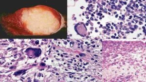
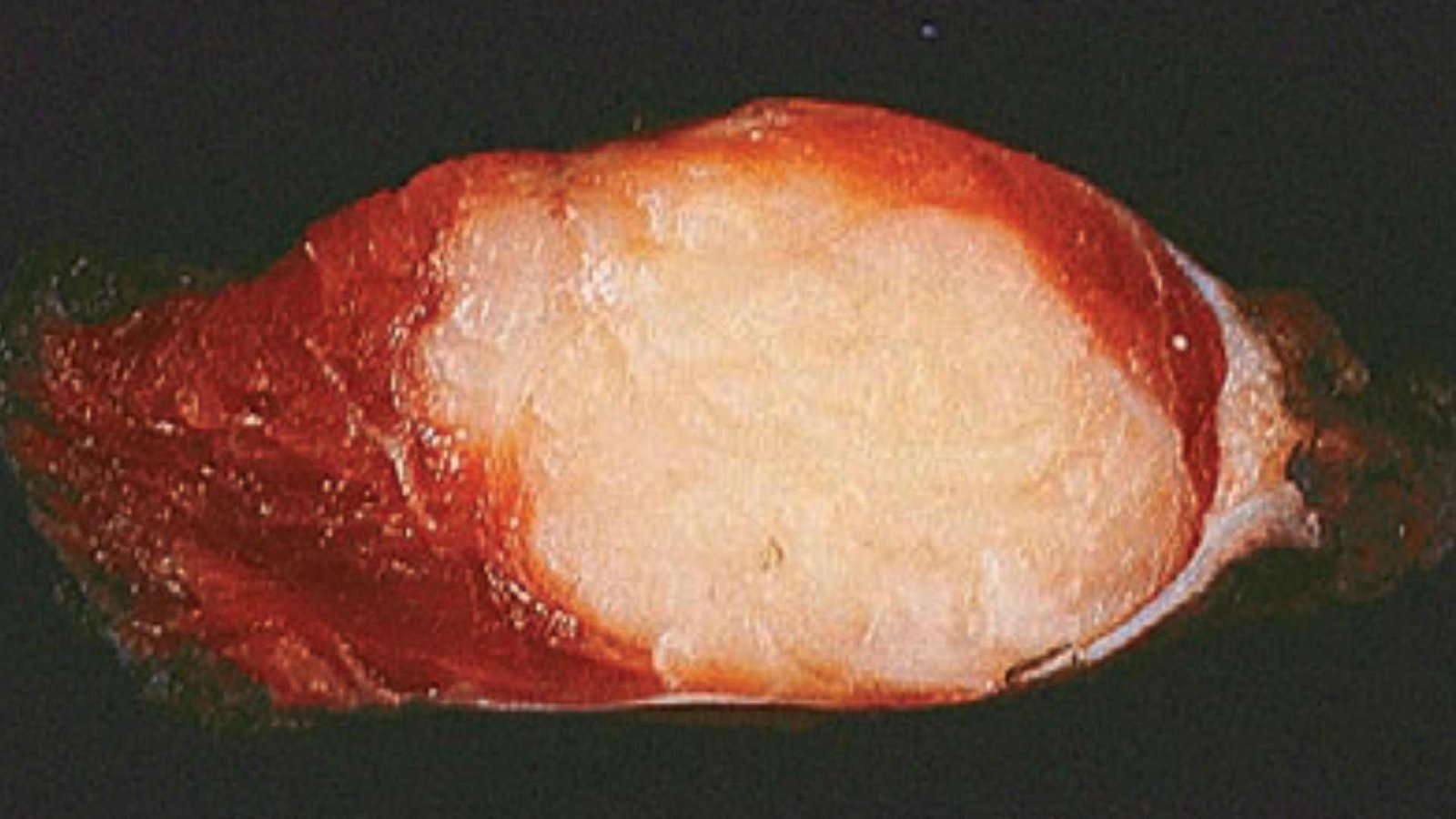
Rhabdomyosarcoma types and morphology
If sarcomas have any kind of alveolar morphology, they should be classified following,
Embryonal Rhabdomyosarcoma
It is a variant & related tumour mass. This sarcoma is most common in children and when growing underneath a mucosal membrane, such as the urinary bladder, vagina, or nasal cavity, etc.
The tumour regularly forms large polypoid masses (soft grey infiltrating stages of differentiation and also consists of primitive round & spindle cells) which resemble a bunch of grape so-called sarcoma botryoides.
The sarcoma botryoides contain rhabdomyoblast with straplike cytoplasm and the characteristic feature is the presence of a dense zone of undifferentiated tumor cells immediately below the epithelium tissue which is called the cambium layer.
Alveolar rhabdomyosarcoma
It is also the most common soft tissue sarcoma of childhood and regularly contains fusions of the FOXO1 gene to either the PAX3 or the PAX7 gene.
PAX3 is a transcription factor that differentiates skeletal muscle and appears as a chimeric PAX3-FOXO1 fusion protein that interferes with differentiation.
It shows a network of fibrous septae and divides the cell into aggregates that resemble pulmonary alveoli. The tumour cells are uniformly round-shaped cells with little cytoplasm and are minimally cohesive in nature.
Pleomorphic rhabdomyosarcoma
Rhabdomyosarcoma in adults is the rarest sarcoma and is commonly seen predominantly in adults (in deep tissues) and characterized by numerous large multinucleated (sometimes), bizarre eosinophilic tumour cells which resemble other pleomorphic sarcomas. Myogenin protein is most commonly seen in this sarcoma.
Causes & symptoms
What causes rhabdomyosarcoma?
Your doctor or consultants do not know the exact causes of most of the rhabdomyosarcoma but some risk factors are associated with it. Some risk factors which cause the cancers are the following,
- Age and gender: RMS is most common in children younger than 20 years, but it can also develop in teenagers and adults. It is relatively more common in boys than in girls.
- Inheritance conditions: Few people have a propensity to develop certain types of sarcoma because they have inherited changes in their DNA from their parents.
Few rare inherited states increase the risk of RMS and normally some other tumours as well: Your doctor explains these few conditions such as,
- Li-Fraumeni syndrome: This syndrome is present in members of families who are more likely to develop sarcomas like RMS and other cancers such as breast cancer, leukaemia, etc.
- Beckwith-Wiedemann syndrome: Children who have this syndrome have a high risk of developing sarcomas like RMS, but they are also more likely to develop Wilm tumour (a type of kidney tumour) and some other cancers.
- Noonan syndrome: It is a condition in which most of the children tend to be short, with heart defects, and can be slower than other normal children in developing physical as well as learning skills. Those children who have this syndrome are also at higher risk for RMS.
- Neurofibromatosis type 1: It is also called von Recklinghausen disease and generally causes multiple nerve tumours (especially in nerves of the skin ), but it also increases the risk of sarcoma of skeletal muscle (RMS).
- Costello syndrome: It is a very rare case present in people but Children with this type of syndrome have high birth weights but then fail to develop well and are short. They also tend to have an enlarged head. They have a chance to develop RMS as well as some other cancers.
- Exposure before birth: Those children who are exposed to x-rays before birth might be linked with a high risk of RMS. Parental use of drugs like marijuana and cocaine has been suggested as a feasible risk factor for RMS.
What are the signs and symptoms of Rhabdomyosarcoma?
Rhabdomyosarcoma can start anywhere in your body, so the symptoms of RMS vary based on location, size, and spread to other parts. They are following rhabdomyosarcoma symptoms,
- In the neck, chest, arm, back, leg, or groin region: swelling or lump, Sometimes pain, redness, or other problems may be seen.
- Around the eye: bulges out of the eye, droopy eyelids, or sometimes the child appears to be cross-eyed, Difficulty in vision.
- In the ear or nasal sinuses: earache, nosebleeds, sinus congestion, headache.
- In the bladder or prostate or vagina: blood in the urine, vaginal bleeding, hard or painful urination, or bowel movements.
- In the abdomen or pelvis: vomiting, nausea, belly pain, or constipation (stool becomes difficult to pass out)
- In bile ducts: RMS is rarely present but some common signs and symptoms like yellowing of the eyes or skin (jaundice).
- Other common symptoms such as bone pain, constant cough, weakness, or weight loss
If you have one or more of these symptoms present then you may go to visit the doctor. Many of these signs and symptoms are more probable to be caused by RMS.
Diagnosis and tests
How is rhabdomyosarcoma diagnosed & tested?
People have some common signs and symptoms that might suggest that a person may have rhabdomyosarcoma (RMS), but tests are needed to find out the disease.
Medical history and physical exam
- Medical history: If your child has signs and symptoms that could be from RMS or (another form of tumour), the doctor or consultant will want to get a complete medical history to find out more about the symptoms present in the child’s patients.
- Physical exam: Doctors take a physical examination to look for possible signs of RMS or other health abnormalities. For example, doctors or consultants find out and feel the lumps or swelling in certain tumours.
Imaging tests: Your doctors or consultants use imaging tests to identify suspicious areas, assess the extent of a tumour, determine how far it has spread, and evaluate the effectiveness of treatments. The tests they use include:
- X-rays (plain X-rays): Plain X-rays are more helpful when looking for soft tissue tumours like RMS.
- CT-Scan (computed tomography): In this test, your doctors inject contrast substances into a vein before the scan to help see more details and better ways.
- MRI (Magnetic resonance imaging) Scan: In this test, your doctors inject contrast material such as gadolinium into your vein before the scan to help see more details about your tumours. It is particularly beneficial if the tumour is located in areas like the head, neck, arm, leg, or pelvis.
- Positron emission tomography (PET) scan: In this test, doctors inject a radioactive substance {Fludeoxyglucose (FDG), which is similar to glucose} into your bloodstream. Cancer cells use more sugar for energy because they grow quickly.
Ultrasound: It uses no radiation and is also helpful for the diagnosis of RMS and other tumours.
Biopsy: Doctors or consultants use a needle to take a sample of tissue from the tumour observed by microscopy and diagnose it with the help of their microscopy features.
Lumbar puncture (spinal tap): Your doctors inserted a small, hollow needle in between the bones of the spine to withdraw some amount of the fluid (CSF), then the sample was sent to the lab for testing and help to diagnose cancer.
Blood Tests: In the RMS blood tests should not be preferred but in some conditions, your doctors prefer the blood test to diagnose cancer.
Management & Treatment
There are various types of treatment available depending on the type of cancer, whether it has spread or not so, where. The types and duration of treatment vary depending on the type of cancer and whether it may be spread. There is the following treatment:
- Surgery: A doctor removes the tumour mass and a few numbers of cells surrounding healthy tissue because it reduces the recurrence of the tumour.
- Radiation therapy: Radiologists use high-power X-rays to help decrease the risk of the tumour coming back again when surgery is undergone. Again and again, people have radiation therapy before surgery, so that the surgeon can remove less amount of tissue. But in the case of RMS radiation therapy is typically given along with chemotherapy.
- Chemotherapy: Anti-cancer drugs kill cancer cells all around the body. Most chemotherapy is delivered injected into a vein or taken by mouth (orally). In some clinical cases, doctors use chemotherapy before surgery because it makes the tumour smaller. Doctors or consultants may recommend chemotherapy to kill any cancer cells left behind after surgery.
- For the low-risk group people, the main combinations of drugs used are:
- VA: Actinomycin-D (vincristine and dactinomycin)
- VAC: vincristine, dactinomycin, and cyclophosphamide
- For the intermediate-risk group people, the most common regimens are:
- VAC: vincristine, dactinomycin, and cyclophosphamide.
- VAC/VI: vincristine, dactinomycin, and cyclophosphamide alternating with vincristine and irinotecan.
What are the complications of rhabdomyosarcoma?
- Hair loss
- Mouth sores
- Loss of appetite
- Nausea and vomiting
- Diarrhea
- Increased chance of infections (few white blood cells preset)
- Easy bruising or bleeding (Few blood platelets present)
- Fatigue (Red blood cells decrease)
Prevention
How can you prevent rhabdomyosarcoma?
RMS cannot be prevented. If you can reduce your risk factors of soft tissue cancers by avoiding long-term exposure to toxic compounds and radiation and changing your lifestyle (staying at a healthy weight or quitting smoking).