What is the knee joint?
The knee joint is a synovial joint of modified hinge variety. Actually, it is a compound joint consisting of three articulations: right and left condylar joints between the condyles of the femur and tibia, and one saddle joint between the femur and patella.
Articular surfaces
- The articular surfaces of the femur of the medial and lateral condyles.
- The trochlear surface of the femur.
- The articular surface of the patella.
- The articular surfaces of the tibia of medial and lateral condyles of the leg.
Factors maintaining the stability of the knee Joint
- The strength and movement of surrounding muscles and tendons.
- Medial and lateral collateral ligaments maintain side-to-side stability.
- Cruciate ligaments maintain anteroposterior stability.
- The Iliotibial tract helps in stabilizing a partly flexed knee.
Ligaments of the knee joint
- Capsular ligament.
- Ligamentum patellae.
- Tibial and fibular collateral ligaments.
- Anterior and posterior cruciate ligaments.
- Medial and lateral menisci.
- Oblique popliteal ligament.
- Arcuate popliteal ligament.
- Transverse ligament.
- Coronary ligaments.
Bursae around the knee joint
There are approximately 12 bursae around the knee joint, four anterior bursae, three lateral bursae, three medial bursae, and two posterior bursae.
Anterior Bursae
- Subcutaneous prepatellar bursa (bursa of housemaid’s knee).
- Subcutaneous infrapatellar bursa.
- Deep infrapatellar bursa.
-
Suprapatellar bursa.
Lateral Bursae
- Bursa between the fibular collateral ligament and tendon of biceps femoris.
- Bursa between the fibular collateral ligament and tendon of popliteus.
- Bursa between the tendon of popliteus and lateral condyle of the femur.
Medial Bursae
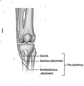
- Bursa, which separates the tendons of sartorius, gracilis, and semitendinosus from each other and from the tibial collateral ligament (bursa anserine).
- Bursa between the tendon of the semimembranosus and medial collateral ligament.
- Bursa between the tendon of the semimembranosus and medial condyle of the tibia.
Posterior Bursae
- The bursa present between the lateral head of the gastrocnemius muscle & the joint capsule in the knee joint.
- The bursa between the medial head of the gastrocnemius and the joint capsule (Brodie bursa).
Relations of the knee joint
- Anteriorly:
- Tendon of the quadriceps femoris, patella, ligamentum patellae, patellar plexus of the nerves, and prepatellar synovial bursa.
- Anteromedially:
- Medial patellar retinaculum.
- Anterolaterally:
- Lateral patellar retinaculum and iliotibial tract.
- Posteriorly:
- Popliteal vessels, tibial nerve, and oblique popliteal ligament.
- Posterolaterally:
- In the upper part, tendon of biceps femoris and common peroneal nerve; in the lower part, lateral head of gastrocnemius and plantaris.
- Posteromedially:
- In the upper part, sartorius, gracilis, semimembranosus, and semitendinosus.
Blood supply
- Five genicular branches of the popliteal artery.
- Descending the genicular branch of the femoral artery.
- Descending branch of the lateral circumflex femoral artery.
- The anterior tibial artery has two recurrent branches.
- A circumflex fibular branch of the posterior tibial artery present in the knee joint.
Nerve supply
- Femoral nerve.
- Tibial and common peroneal nerves.
- Obturator nerve (posterior division).
Movements
- Flexion.
- Extension.
- Medial rotation.
- lateral rotation
Locking of the knee
- Change in the shape of the articular surface from rounded to flat increasing the area of articulation.
- Medial rotation of femur tightening all the associated ligaments.
- Line of gravity passing anterior to the joint.
- Popliteus unlocks the joint by rotating the femur laterally.
Unlocking of the knee joint
- Lateral rotation of the femur on the tibia during the initial phase of the flexion.
- It is brought about by the popliteus muscle.
- The unlocked knee can be further flexed.
- All ligaments are relaxed.
Applied anatomy
- Osteoarthritis
- Injuries to cruciate ligaments
- Aspiration of the knee joint
- Arthroscopy of the knee joint
- Knee replacement
- An unhappy triad of the knee joint
- Housemaid’s knee
- Clergyman’s knee
- Baker’s cyst
- Meniscal tears
[embeddoc url=”https://notesmed.com/wp-content/uploads/2020/09/Knee-joint-1.pdf” download=”all”]