About Hymenolepis nana
The common name of Hymenolepis nana: The Dwarf tapeworm
History and Distribution
- Hymenolepis means a thin membrane covering the egg (Greek hymen—membrane, lepis—rind or covering), and nana means small size (nanus—dwarf).
- 1st discovered by Bilharz in 1857
- H. nana is the smallest intestinal cestodes that infecting to humans beings.
Epidemiology
- Cosmopolitan in distribution but is more common in a warm climate than in cold climates.
- Infection is most common in institutional populations and school children.
- Common parasite of mice.
Habitat: Small intestine (proximal ileum of man), rodents like mice and rats (found in the posterior part of the ileum).
Morphology
- Adult Worm
- Eggs
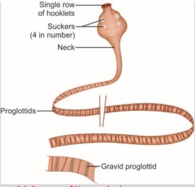
Adult Worm:

- Hymenolepis nana is the smallest intestinal cestode that infecting to humans being.
- Around 5 to 45 mm in length and less than 1 mm thick.
- Scolex: It is globular with 4 suckers, & a retractile rostellum with a single row of hooklets(20-30), rostellum remains invaginated in the apex of an organ.
- Neck: long slender.
- Strobila:
- Generally, it’s consisting of 200 or more proglottids, which are much broader than long.
- Segment-0.3×0.9 mm.
- Genital pores are marginal on the same side.
- The testis is round and 3 in number and the Uterus has lobulated walls.
- Eggs are released in the intestine by the disintegration of the distal gravid segments.
Egg:

- Roughly spherical or ovoid in shape.
- 30–45 μm in size in diameter.
- It contains two distinct membranes:
- The outer membrane is thin colorless.
- Inner embryophore enclosing the hexacanth oncosphere with three pairs of hooklets.
- The space between the 2 membranes is filled with yolk granules and 4–8 thread-like polar filaments arising from 2 knobs on the embryophore.
- The eggs float in a saturated solution of salt.
- Non-bile stained.
- Instantly infective & unable to survive for more than 10 days in the external environment.
Mode of infection:
- Ingestion of food contaminated with eggs.
- Autoinfection: External and internal
- External autoinfection occurs when a person ingests their own eggs by the fecal-oral route
- Internal autoinfection: occur when the eggs released in the intestine hatch there themselves.
- Rarely by ingestion of food contaminated with fleas harboring the cysticercoid larvae.
Life Cycle of Hymenolepis nana

- Definitive host: Man
- No intermediate host
- Infective stage: embryonated egg
- Direct
- Indirect
- Autooinfection (Internal, external)
Steps
- H. nana is uncommon in that it undergoes multiplication in the body of the definitive host.
- When a large number of eggs are swallowed, or in internal autoinfection, they hatch in the small intestine.
- When the hexacanth embryo penetrates the intestinal villus and then develops into the cysticercoid larva.
- It is a solid pyriform structure.
- A solid pyriform contain the vesicular anterior end which containing the invaginated scolex and a short conical posterior end.
- After about 4 days, the mature larva emerging out of the villus and then evaginates its scolex and attaches to the mucosae.
- Now it’s starts strobilization, to become the mature worm, which begins producing eggs in about 25 days.
- A different strain of H. nana infects rats and mice.
- The eggs passed in rodent feces are ingested by rat fleas (Xenopsylla cheopis and others), which acts as the intermediate host.
- The eggs develop into cysticercoid larvae in the hemocoel of these insects.
- Rodents get infected when they eat these insects.
- The murine strain does not appear or arrive to infect man.
- However, the human strain may infect rodents, which may, therefore, constitute
Clinical features:
Generally asymptomatic
- Abdominal pain
- Diarrhea
- Nausea
- Pruritus-sometimes
Laboratory diagnosis:
- Direct microscopy: demonstration of characteristic eggs in feces.
- Concentration methods: like salt flotation and formalin ether may be used.
- Serological methods: ELISA test: 80% sensitivity.
Treatment:
- Niclosamide: 60-80 mg/kg for 5-7 days.
- Praziquantel: 25 mg/kg in a single dose at one time (acts both against the adult worms and the cysticercoids in the intestinal villi).
Prophylaxis:
- Maintenance of good personal hygiene.
- Maintenance of sanitary improvements.
- Avoid contaminated food and water.
- Rodent control.