What are Gallstones disease (Symptomatic Cholelithiasis)?
Gallstone disease (symptomatic cholelithiasis) is one of the most common biliary pathology and surgical problem in the world.1,2 Cholelithiasis is hard, crystal-like deposition that can form in your gallbladder which lies in your organ called the liver.3
Its size ranges vary from as small grains like sand or millet to as large like golf balls-however small stones are most common.1, 2, 14 The rises in gallstone (cholelithiasis) disease freight and a broad spectrum of non-specific presentation make the disease more difficult.1
Worldwide scheme the incidence of gallstones illness ranges from 3 % to 21.9 %. The incidence of gallstones in Asia is 4% to15%.3,4 This gallstones disease (symptomatic cholelithiasis or non-symptomatic cholelithiasis) is most commonly present in females with a ratio of female to man: (4:1).5,6
Keywords: Gall Stones; Common Bile Duct; Gallstone Disease (symptomatic Cholelithiasis).
What are the types of Gallstones?
There are the following types of gallstones;2, 14
- Cholesterol stones (6 %): solitary stone
- Mixed stones (90 %): Cholesterol compounds, Salt of calcium with phosphate carbonate, palmitate compound, various proteins, and multiple faceted.
- Pigment stones (4 %): small, black or greenish-black, multiple.

What are the Symptoms of gallstone disease (symptomatic cholelithiasis)?
The majority of cases of gallstone disease are asymptomatic (around >80%). Approximately 1% to 2% of asymptomatic patients will have to develop various symptoms that require proper surgery every year.2
Symptoms typically occur when gallstones cause your gallbladder spasm or bile duct obstructions. In these cases, There are following manifestations occur:
Abrogate pain in the upper portion of the right or middle part of your abdomen.
Fever or chills.
Nausea and Vomiting.
Yellowing of the eyes or skin (jaundice).
Itchiness.
Dyspepsia.
Flatulence.
Food intolerance like fat.
Alteration of bowel frequency.
Dark color urine.
Light-colored stools.
Where is the pain located?
The main symptom of gallstones disease (symptomatic cholelithiasis) is pain in the right upper quadrant or middle (epigastric pain) part of the abdomen.
It is directly below the ribs, which may radiate to the backside and also sometimes can spread to the arm, right shoulder, or chest.2, 14
Some patients felt the pain as dull aching pain or sharp and stinging deep pain. The pain is also described as colic or colicky pain and it is aggravated by a heavy meal that starts during the night and wakes the patient.2. 14
The gallstones pain is sometimes more confused with the pain of heart attack. The pain generally lasts between the last few minutes to the last few hours.2, 14
What are the causes of the gallstones disease (symptomatic cholelithiasis)?
In general, gallstones formed from there is a variation in the composition of bile. Bile formation occurs in your organ liver and is stored in your gallbladder, and then finally delivered into the intestine.2, 7, 14
The majority of the stones are formed from cholesterol and are called cholesterol stones. When the amount of cholesterol within the bile is too high and causes the formation of the solid ball-like stone.2, 7, 14
It is less common, formed from pigment bilirubin, which is formed from the red blood cells (RBC) breakdown product and also gallstones create by a disproportion of bile salts, lecithin compound, or calcium carbonate.2, 7, 14
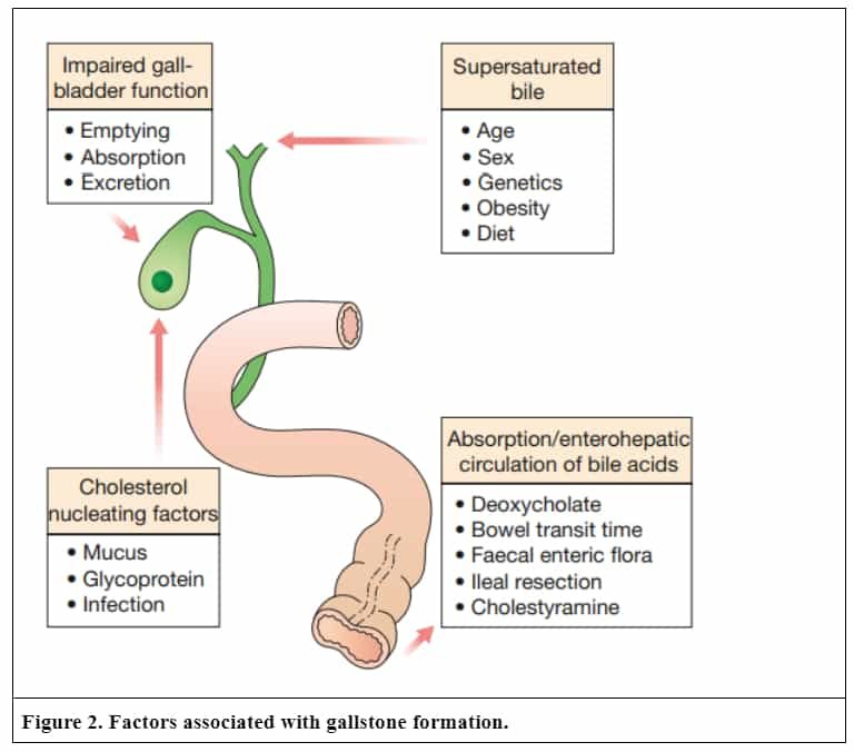
Altered Gallbladder function such as stasis (estrogen therapy, vagotomy, pregnancy, and long-term intravenous fluids), poor emptying, poor absorption and some infection (E.coli, Salmonella, Ascaris lumbricoides, Clonorchis sinensis).2, 14
Supersaturated bile due female, fertile, fat, forty, and high-calorie diet.2, 14
Altered enterohepatic circulation due to ileal resection, ileal diseases, altered bowel transit time, altered bowel flora, cholestyramine, and deoxycholate.2, 14

Risk Factors
The most common risk factors for the formation of gallstones are:2,14
Female, fertile, fat, forty.
Obesity or overweight.
Pregnancy and age over 40 years old.
Family members with gallstones.
Some medications (Estrogen therapy).
Geography such as European, Native American, or Hispanic descent.
Some clinical conditions such as diabetes, sickle cell anemia, cirrhosis of the liver, cystic fibrosis, thalassemia, malaria, and inflammatory bowel disease (like Crohn’s disease).
What is the diagnosis for Gallstones disease (Symptomatic Cholelithiasis)?
In your doctor or consultant, take a thorough medical history and take out a thorough physical examination.
A doctor or examiner can press on your upper right quadrant or right part of the abdomen and ask the patient to take inspiration (breath) deeply during the examination the patient experiences pain that may indicate an inflammation of the gallbladder (Murphy’s sign).10
There is the most common test which can help to diagnose
- Blood test: Total WBC count (leukocytosis).
- Liver function test: Elevated total bilirubin, alkaline phosphatase, etc.
- Plain X-ray: Only 10% of gallstones are radio-opaque.
Abdominal Ultrasound
- It is a rapid non-invasive diagnostic test that uses sound waves to confirm the gallstones’ presence or absence and inflammation of the gallbladder.
- Gallstones are seen with posterior.
Endosonography:9
- It consists of a flexible tube and at the end of the tube, the ultrasound probe is attached.
- First, insert the flexible tube into your mouth and then pass it forward into the patient’s abdomen and capture the ultrasound image.
Endoscopic retrograde cholangiopancreatography (ERCP)10, 11, 12, 13, 14
- It is the most preferable method and using an endoscope if a stone is suspected to block your bile duct.
- A doctor moves the endoscope through the person’s mouth to the throat to the esophagus and finally reaches into your stomach and upper intestine.
- A doctor should visualize patients’ bile duct using some special contrast media and if possible, sometimes the stone is removed.
CT -Scan 2, 14
- This is an imaging procedure that helps to confirm the presence of gallstones in your bile duct and also detect possible complications, like obstruction of the bile duct and pancreatitis.
- Magnetic Resonance Imaging (MRI)
- It is a 3D imaging procedure utilizing a strong kind of magnetic field to assemble
- the detailed structure in your body.2, 14
How are your gallstones disease (Symptomatic Cholelithiasis) treated?
In general, gallstones are usually only treated for underlying causes. There are a few general measures are presented that help to improve your health:2, 9, 10, 11, 12, 13
Decreasing intakes of fatty foods.
Temporary fasting of food.
Administration of medications that help to reduce body spasmodic tension (pain) against the blockade caused by the stone.
Administration of painkillers that help to reduce pain.
Other various methods depending on the gallstone’s exact location in your gallbladder.
Surgically to remove your gallstones
The surgical process helps to remove the gallbladder, which is known as cholecystectomy. It is the most preferred treatment for symptomatic cholelithiasis (gallstones with symptoms) i.e. when gallstones are present with severe discomfort.
It is the most common, reliable, authentic, and most effective procedure to eradicate symptoms in the long term and to prevent further possible complications.9,10
There are two types of surgical techniques for removing your gallbladder.
Laparoscopic cholecystectomy (LapChole)
LapChole is a keyhole type of minimally invasive surgical method, we used for the removal of the gallbladder disease and its various complication.2, 10, 14
It is typically operated on for treatment of acute cholecystitis or chronic cholecystitis, symptomatic cholelithiasis, acalculous cholecystitis, biliary dyskinesia, pancreatitis gallstone, and gallbladder polyps or masses.2, 10, 14
Open Cholecystectomy:
It is an older version of surgery that requires a larger incision in your abdomen to extract your gallbladder. This type of surgery requires more time for recovery.8, 10
It is done through right-sided subcostal Kocher’s incision when used if a patient is not fit for laparoscopic surgery (LapChole); Suspected gallbladder carcinoma; in suspected CBD stones; Mirizzi syndrome.
After Surgery of the gallbladder
When people show normal lives as well as healthy lives without a gallbladder. After the withdrawal of the gallbladder.9, 10 The bile flows directly from your liver into your intestine via the connecting duct called the bile duct with stored in your gallbladder.9, 10
And bile always helps to support the digestion of food as usual but in some cases, mild diarrhea or different digestive disorders may occur. The risk of complications from cholecystectomy (removal of the gallbladder) is might be low.9, 10
Endoscopic retrograde cholangiopancreatography (ERCP):
ERCP is one of the major intrusive endoscopic techniques employing x-rays and special contrast media (dye) to envisioned biliary and pancreatic ducts.2, 10, 14
It is generally used for both diagnostic and therapeutic propose.2, 10, 14 If there are further gallstones in the gallbladder, your doctor recommended surgery.
Magnetic resonance cholangiopancreatography (MRCP):
MRCP is a Magnetic Resonance Imaging (MRI) analysis of a noninvasive visualization of biliary ducts and pancreatic ducts.2, 10, 14
Non-surgical treatments Procedure:
Non-surgical treatments are normally suggested if surgery is not feasible.
There are the following treatments alternatives:
Extracorporeal shock wave lithotripsy (ESWL):
ESWL is a nonsurgical alternative approach to address gallstones. It is admiringly efficacious for stones of the bile duct in patients in whom endoscopic or surgical stone removal fails.11 The side effect is lung damage but there are no other significant side effects.11
Medications
Medical dissolution therapy with bile acids is the alternative method for patients with various (mild-to-moderate) symptoms due to cholesterol gallstones.2, 12, 14
Chenodeoxycholic acid (CDCA, chenodiol) drug has been chiefly replaced by the safer and more efficient medication ursodeoxycholic acid (UDCA) but it is not very successfull.2, 12, 14
What is the prognosis for Gallstones disease (symptomatic Cholelithiasis)?
In the case of gallstone disease (symptomatic cholelithiasis) do not cause any sign and symptoms and don’t require any treatment.14 However signs and symptoms occur, treatment is essential. However, the general prognosis of gallstones is awesome and most of the patients fully recover.14
What are the complications of Cholelithiasis (Gallstones)?
In the case of gallstone disease (symptomatic cholelithiasis), there are the following complications based on-site or organs:15
In the gallbladder.2, 15
Silent asymptomatic stones (10% of males and 20% of females)
Biliary colic
It is severe within hours after a meal (fatty/heavy meal).
Acute cholecystitis
Acute inflammation of the gallbladder.
Chronic cholecystitis
Chronic inflammation of the gallbladder.
Empyema gallbladder.
Perforation
It causes biliary peritonitis or pericholecystic abscess.
Mucocele of gallbladder.
Limey gallbladder.
Gallbladder carcinoma.
In the CBD15
Secondary CBD stones,
It occurs in 10% of gallstones.
Cholangitis
Inflammation of bile duct.
Pancreatitis
Inflammation of parenchyma of the pancreas.
Mirizzi syndrome:
Due to squeeze of common hepatic duct (CHD) / common bile duct (CBD) by stone from a cystic duct or cholecysto-choledochal fistula.
In the Intestine
Cholecystoduodenal fistula operates gallstone ileus and obstruction of the intestine.15
If these complications are present in your body they often need emergency treatment.
REFERENCES
- Clinical Profile of Patients Presenting with Gallstone Disease in University Hospital of Nepal. [PubMed | Full Text]
- Bailey and Love’s Short Practice of Surgery-27th edition
- Everhart JE, Khare M, Hill M, Maurer KR. Prevalence and ethnic differences in gallbladder disease in the United States. Gastroenterology. 1999 Sep 1;117(3):632-9.
- Stinton LM, Shaffer EA. Epidemiology of gallbladder disease: cholelithiasis and cancer. Gut and liver. 2012 Apr;6(2):172.
- Singh V, Trikha B, Nain C, Singh K, Bose S. Epidemiology of gallstone disease in Chandigarh: A community‐based study. Journal of gastroenterology and hepatology. 2001 May;16(5):560-3.
- Idris SA, Shalayel MH, Elsiddig KE, Hamza AA, Hafiz MM. Prevalence of different types of gallstone in relation to age in Sudan. Sch. J. App. Med. Sci. 2013;1(6):664-7.
- AMBOSS. “Cholelithiasis, choledocholithiasis, cholecystitis, and cholangitis.” November 28, 2018. Accessed June 19, 2019.
- Medscape. “Gallstones (Cholelithiasis) Treatment & Management.” March 30, 2017. Accessed June 19, 2019.
- BMJ Best Practice. “Cholelithiasis.” January, 2018. Accessed June 19, 2019.
- Hassler KR, Collins JT, Philip K, Jones MW. Laparoscopic Cholecystectomy. 2021 Sep 28. In: StatPearls [Internet]. Treasure Island (FL): StatPearls Publishing; 2022 Jan–. [PubMed]
- Paumgartner G, Sauter GH. Extracorporeal shock wave lithotripsy of gallstones: 20th anniversary of the first treatment. Eur J Gastroenterol Hepatol. 2005 May;17(5):525-7. [PubMed | Full Text | DOI]
- Konikoff FM. Gallstones – approach to medical management. MedGenMed. 2003 Oct 15;5(4):8. [PubMed]
- Friedman GD, Raviola CA, Fireman B. Prognosis of gallstones with mild or no symptoms: 25 years of follow-up in a health maintenance organization. J Clin Epidemiol. 1989;42(2):127-36 [PubMed | Full Text | DOI]
- SRBs Manual of Surgery-5th edition.