What is the cerebellum?
The cerebellum is the largest part of the hindbrain weight is about 150 gram situated dorsal to the pons and medulla and separated from them by the fourth ventricle.
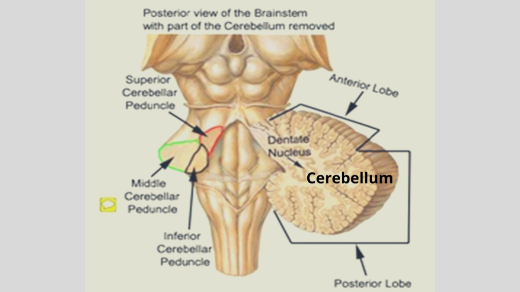
The cerebellum connected to the brainstem by the following pair structures;
- Superior cerebellar peduncles: the midbrain.
- Middle cerebellar peduncle: pons.
- Inferior cerebellar peduncle: medulla oblongata.
The cerebellum is occupied posterior cranial fossa in the skull, where it is covered by tentorium cerebelli and present and beyond the pons and medulla oblongata in the brain.
Basic Functions of the cerebellum
- Maintenance of equilibrium.
- Regulation of muscle tone.
- Coordination of somatic motor activities.
Gross anatomy of the cerebellum
It consists of inner white matter and outer grey matter with 4 pairs of deep cerebellar nuclei. The outer cortex is thrown into numerous transverse folds called folia which are separated by fissures.
Each folium contains a core of white matter which is covered superficially by gray matter in the cerebellum.
The central core of the white matter is arranged in the form of the branching pattern of a tree, which is called arborvitae cerebelli (arborvitae = tree of life).
Intracerebellar Nuclei
It also called central nuclei. They are masses of grey matter which is embedded in the white matter of the cerebellum. From lateral to medial, these nuclei are:
- The dentate
- The emboliform
- The globose, and
- The fastigial.
Anatomical subdivisions
It consists of 2 cerebellar hemispheres & which are joined by a narrow median vermis in the cerebellum. It is divided into the following three lobes:
- The anterior lobe
- The middle lobe/ posterior
- The flocculonodular lobe
- It separated by primary and posterolateral fissure/uvulonodular fissures.
- Horizontal fissure:
- It separates the superior surface from the inferior surfaces. This fissure is most conspicuous (noticeable) and runs along the lateral and posterior margins of the cerebellum.
The inferior vermis is separated from the hemisphere by vallecula.
Morphological subdivisions
Archicerebellum
- First to appear in evolution in aquatic vertebrates
- Includes floculonodular lobe & lingulla
- Receives input from vestibular nerve & vestibular nuclei.
- Concerned with the maintenance of body equilibrium, and posture of trunk muscles.
Paleocerebellum
- Appears next in terrestrial vertebrates.
- Includes anterior lobe except for lingula & the pyramid & uvula.
- Receives proprioceptive & exteroceptive impulses from the spinocerebellar & Cuneo cerebellar tract.
- Role in the maintenance of tone of voluntary muscle posture and crude movements of the lower limb.
Neocerebellum
- Last to appear with the appearance of neocerebrum.
- Includes middle lobe except for pyramid & uvula.
- Has extensive connection with the cerebral cortex through pontine nuclei.
- Concerned with the regulation of fine and skill voluntary movements.
Microanatomy of the cerebellum
It consists of 3 layers:
- Molecular – outer
- Purkinje – middle
- Granular layer- inner
Molecular (Plexiform) layer:
- Contains the outer stellate cell and the inner basket cell & large numbers of unmyelinated fibers.
Purkinje Cell Layer:
- It is flask-shaped with arranged in a single layer.
- Dendrites of these cells which passes into the molecular layer & undertake profuse branching.
- Axons which is passes throughout the granular layer to enter the white matter.
- The synapse with cells of one of the intracerebellar nuclei present in this layer.
Granular layer:
- It contains granular cells & Golgi cells in this layer.
- The dendrites of the granular cells which is synapse with the mossy fibers.
- The axon passes into the molecular layer of the cerebellum, where it bifurcates at a T junction.
White matter
Three groups of fibers:
- Intrinsic:
- It connects different regions of the cerebellum of the same side or two cerebellar hemispheres.
- Afferent:
- The afferent fibers enter the cerebellum mainly through the inferior cerebellar peduncles and middle cerebellar peduncles.
- Efferent:
- It is formed by the axons of the Purkinje cells which pass & synapse deep cerebellar nuclei in the cerebellum.
- The efferent fibers from dentate, emboliform, and globose pass throughout the superior cerebellar peduncle.
- The efferent fibers from the fastigial nucleus that leave throughout the inferior cerebellar peduncle.
Blood supply of the cerebellum
- The superior cerebellar artery:
- It is a branch of the basilar artery that supplies the superior surface of the cerebellum in the brain.
- The anterior inferior cerebellar artery:
- It is a branch of the basilar artery that supplies the anterior part of the inferior surface of the cerebellum in the brain.
- The posterior inferior cerebellar artery:
- It is a branch of the vertebral artery that supplies the posterior part of the inferior surface of the cerebellum the brain.
Sign & symptoms of cerebellar disease
- Hypotonia
- Alteration of gait/ lurching gait
- Ataxia– intentional tremor
- Dysdiadochokinesia– inability to perform the regular alternating movement of the body
- Dysmetria
- Dysarthria or scanning speech, i.e., speech is slurred, monotonous with pauses at wrong places.
[embeddoc url=”https://notesmed.com/wp-content/uploads/2020/10/Cerebellum.pdf” download=”all”]