Morphology of Mycobacterium leprae
Mycobacterium leprae is straight rods about 1 – 8 x 0.2 – 0.5µm and it appears either single or groups. It is an intracellular low acid-fast bacillus with 5% H2SO4. It is bound together like cigar bundles by a lipid-like substance called Glia. The Globi present in Virchow’s lepra cells or Foamy cells.
Cultivations: The generation time is around 12 to 12 days and the methods of cultivations are the footpad of mice and the nine-banded armadillo.
Pathogenesis of Mycobacterium leprae
At first, bacilli enter through the respiratory tract and peripheral nerve such as Schwann cells in cooler places (cutaneous nerve and peripheral nerve trunks of limbs and face), and then bacilli multiply in the Schwann cells. After multiplication, they go to either good or weak cell-mediated immune(CMI) response. In the good CMI response, there is no skin/nerve lesion appear, or skin and nerve lesion appear followed by spontaneous healing or paucibacillary (PB) leprosy. And in the weak CMI response cases, there is multibacillary leprosy condition with addition to skin and nerve, eyes, testes, kidney, voluntary/smooth muscles, reticuloendothelial system, and vascular endothelium get involved. Finally, it goes to disabilities and deformities.
Laboratory Diagnosis of leprosy (Mycobacterium leprae)
Specimen collection
In the case of leprosy, there is a total of six samples are collected from different places such as four from the skin (forehead, cheek, chin, and buttock), one from the ear lobe, and one from the nasal mucosa by nasal scraping/blow.
Slit skin smear
It is a technique followed to collect the sample from the skin and ear lobes. The preferred site is an edge of the lesion and the lesion is cleaned with spirit, then it is pinched uptight to minimize bleeding.
A scalpel is made a 5 mm long incision, deep enough to get into the infiltrate layers. After wiping off lymph or blood that may have exuded, transversely rotated the scalpel blade to scrape the sides and base of the incision so as to obtain a tissue pulp from below the epidermis and which is smeared uniformly over an area on a slide have 8 mm diameter.
Nasal blow
In this method, the early morning mucus sample is collected by blowing the patient’s nose on a clean cellophane sheet.
Nasal scraping
In this method, using a mucosal scraper to scrape the nasal septum sufficiently so as to remove a piece of a mucous membrane, and transferred it onto a slide and which teased out into a smear uniformly.
Microscopy
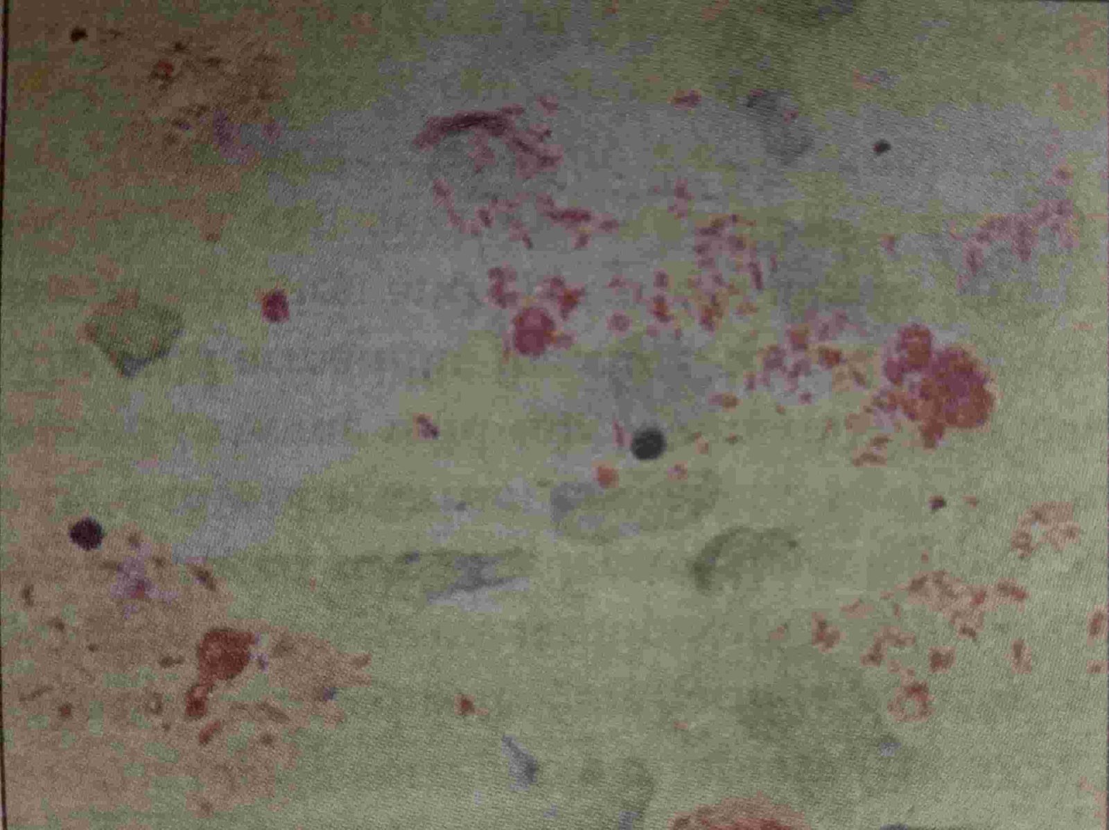
The smear microscopy is done to demonstrate the low acid-fast bacilli in the lesion by using the Ziehl-Neelsen technique by using 5% sulfuric acid for decolorization.
Under the oil immersion field, red acid-fast bacilli arranged single or cigar-like bundles (in groups) and bonded together by a lipid-like substance, the glia to form globi.
The globi are present inside the foamy macrophages which are called Virchow’s lepra cells or foamy cells.
Live bacilli are uniformly stained with parallel sides and around ends, solid, and length is five times the width.
Dead bacilli have a fragmented and granular appearance and less uniformly stained.
Bacteriological index (BI)
BI is based on the total number (dead or live) of bacilli seen per oil immersion field(OIF). It helps to calculated by totaling the number of pluses scored in all the smears per unit the number of smears examined.
Morphological index (MI)
MI is expressed as the percentage of uniformly stained bacilli per unit of the total number of bacilli counted. It is a better marker to monitor the treatment response. During clinical treatment, the MI should fall down whereas the BI may remain constant.
SFG percentage (solid, fragmented granular rod percentage)
The percentage of solid, fragmented granular rods are recorded separately, and this method gives a better picture of bacterial morphology and is a more sensitive indicator of monitoring the treatment response than Ml.
Culture of Mycobacterium leprae
Mouse footpad cultivation: It is not cultivable either in artificial or tissue culture method. They only cultivate M. leprae is by inoculating the specimens intradermally into footpads and keep in at 20 degrees Celcius for 6-9 months.
Other animals such as Nine-banded Armadillo are highly susceptible and Chimpanzees, Mangabey monkeys also are used for culture.
Antibody detection
- FLA-ABS (Fluorescent leprosy antibody absorption test): 92% sensitive and 100 % specific.
- ELISA: It is used to detecting IgM antibodies to phenolic glycolipid-1 (PGL-1) antigen of M.leprae present in 95% of patients with untreated LL patients and the titer decreases with effective therapy. In the TT patients are sensitivity is low around 60%.
Lepromin test
It is a test used for a known prognosis but not a diagnosis. It is discovered by Mitsuda in 1919. It shows the type four hypersensitivity reaction (delayed hypersensitivity reaction) and an intact CMI against the lepra antigen.
The extract of M.leprae is 0.1 mL lepramin antigen is injected intradermally to inner forearm and induration is observed 48 hours later.
- Early reading or Fernandez reaction
- Erythema is produced at the site of inoculation within 48 hours. It corresponds to that of the tuberculin test and indicated a type four hypersensitivity reaction (delayed hypersensitivity reaction) to the soluble lepra antigens.
- The induration is more than 10 mm diameter indicates a positive test, which indicates past exposure to lepra bacilli and it does not indicate active infection.
- Late reading or Mitsuda reaction
- It occurs at three weeks and characterized by the presence of granulomas. It is positive in tuberculoid patients, negative in lepromatous patients.
- The positive test is indicated by a nodule formation with more than 5 mm size at the site of inoculation and which ulcerates later on.
Uses of Lepromin test
- Lepromin test used to classify the lesions of leprosy patients
- Lepromin test used to assess the prognosis and response to treatment
- Assessing resistance to leprosy in individuals
Lucio’s reaction
It is an unusual reaction and seen in some cases of the Caribbean and Mexico. In this reaction, diffuse dermal infiltration occurs without skin lesions so-called diffuse lepromatosis. The Mitsuda test negative.
Management
- Combination chemotherapy: dapsone, rifampin, and clofazimine.
- Antiinflammatory for reactions: steroids, thalidomide
- Supportive care, eyes, hands, feet (to prevent tissue injury related to the neurologic deficit)
WHO recommended Multidrug therapy
The drugs should be used only after a doctor or consultant prescription.
- Paucibacillary case
- Rifampicin 600 mg/ month for 6 months
- Dapsone 100mg / day for 6 months
- For a patient with a single lesion a single dose of Rifampicin (600-mg), Ofloxacin (400-mg), & Minocycline (100-mg)
- Multi bacillary case
- Rifampicin 600 mg per month for 2 or more years
- Dapsone 100 mg/day for 2 or more years
- Clofazimine 300 mg / month + 50 mg / day for 2 or more years.
[embeddoc url=”https://notesmed.com/wp-content/uploads/2021/05/leprosy.pdf” download=”all” text=”Complete pdf file-Download”]