Introduction of Cytomegalovirus
Cytomegalovirus is the largest virus in the Herpesviridae family. It causes massive or huge enlargement of infected host cells.
Morphology
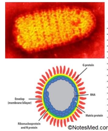
It is large (about 150-200 nm size), spherical in shape with icosahedral symmetry. The nucleocapsid: dsDNA is the largest among herpes viruses, which consist of 240kbp nucleotide. An envelope is of a lipoprotein nature. The tegument is lies between the capsid and envelop and these viruses are strictly species-specific.
Cell type specificity:
- In vivo: Cytomegalovirus infects kidney and salivary glands; where it undergoes latency.
- In vitro: Replicate only human fibroblast cell line and produces a characteristic cytopathic effect (CPE) described as Owl’s eye appearance.
Cell to cell spread: closely associated with cells but very little virus may be cell-free.
Clinical manifestations
- Congenital infections.
- Perinatal infections.
- CMV mononucleosis in adults.
- Severe infection in immunocompromised and transplant recipients.
Epidemiology
Transmission:
- Close person-to-person contact.
- Oral and respiratory spread is the predominant mode.
- Transplacental route (transmission from mother to fetus).
- Blood transfusion: about 0.1-10 %.
- Organ transplantation.
- Sexual contact.
Reservoir: Only humans are the known host for CMV.
Source: urine, saliva, semen, breast milk, and cervical secretions, & is carried in circulating white blood cells in the body.
Endemic: Worldwide present throughout the year without any seasonal variation.
Risk factors: Low socioeconomic status and poor personal hygiene facilitate the infection.
Prevalence is high in underdeveloped countries with 90% of people being seropositive in contrast to 40-70% seropositivity in developed countries.
Laboratory diagnosis of Cytomegalovirus
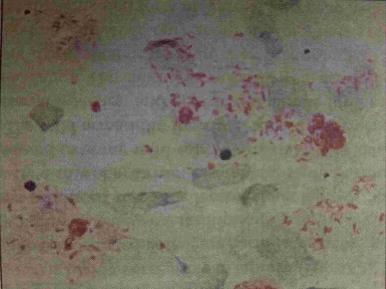
Detection of Inclusion Bodies:
In urine: Produce characteristic perinuclear cytoplasmic inclusions in addition to the usual intranuclear inclusions seen (Owl’s eye appearance).
Virus isolation:
Isolated from throat and urine.
Human fibroblasts are the most ideal cell lines, specific for CMV.
Cytopathic effect: After 2-3 weeks of incubation, the following CPE observed, Typical CMV inclusions and multinucleated giant cells are seen.
Enlargement of infected host cells. Shell vial technique can be followed for early growth detection or determine for about 1-2 days.
Very useful in CMV mononucleosis where viral load is low and CPE takes several weeks to appear.
Antibody detection:
- ELISA.
- Rapid test formates.
Antigen detection: pp65 antigen.
Molecular methods: PCR and real-time PCR.
Treatment
It does not respond to Acyclovir.
- Ganciclovir given IV routes.
- Valganciclovir: Prodrug of ganciclovir given orally.
- Foscamet: effective against ganciclovir resistant.
- Cidofovir: alternate to ganciclovir.
- CMV immunoglobulin: given with ganciclovir for treatment of CMV infections in bone marrow transplant recipients.
Prophylaxis
- Both ganciclovir and valganciclovir have been used successfully for prophylaxis and pre-emptive therapy in transplant recipients.
- CMV immunoglobulin has shown to be effective in preventing congenital infection when given to mothers during pregnancy.
[embeddoc url=”https://notesmed.com/wp-content/uploads/2020/08/Cytomegalovirus.pdf” download=”all”]