Introduction of Adenovirus
Rowe and associates in 1953. The adenovirus is isolated from adenoids originally. Adenovirus shares a common complement-fixing antigen. The virus infects humans, birds, and animals, and infections are most common in children.
Morphology of Adenovirus
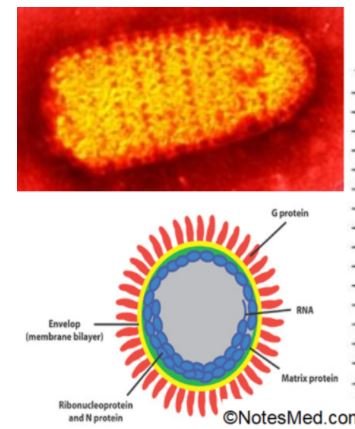
It is a non-enveloped, icosahedral symmetry, linear Double-stranded DNA virus. The size of a virus is 70-90nm and contains 252 capsomers and 240 hexagons. The virus has 12 fibrillar pentons, and it was space vehicle shaped appearance.
Resistance
It is a heat-stable virus and readily inactivated at 50◦C. They resist ether and bile salts.
Epidemiology
It is an endemic disease and is transmitted through droplets, Direct contact, Feco-oral transmission. Its 1/3rd of human serotypes cause human illness.
Virus effects on cells:
It saws in marked rounding, enlargement, and aggregation of affected cells which are grape-like clusters. Rounded intra-nuclear inclusion containing DNA present in the cells.
Pathogenesis
- It generally causes infection of the respiratory tract, eye, GIT & UTI.
- Infection occurs through conjunctiva or nasal mucosa.
- Children- fecal-oral transmission.
- Incubation period: 5-7 days
- Multiply initially in the conjunctiva, pharynx, or small intestine and spread to draining L.nodes
- Serotype 1- 8 most common
- Subgenus(SPS) C – Acute febrile pharyngitis
- Subgenus B- Acute respiratory disease
- Serotype 40 and 41 – infantile gastroenteritis
- Serotype- 8, 19, 37- eye infection
- Serotype 19,37- genital infection
- Serotype 3, 4, 11 – acute follicular conjunctivitis
Immunity
- Induces long-lasting immunity
- Maternal antibodies protect infant
Lab Diagnosis of Adenovirus
Specimen
- Throat swab
- Nasopharyngeal aspirates
- Bronchial lavage
- Conjunctival swab
- Corneal scraping
- Urine
- An anal swab
- Genital secretions
- Feces, rectal swab, and biopsy
Microscopy
- Virus particles in stool by EM
- Virus isolation:
- Primary human embryonic kidney cell line and A549 cell line.
- HEp-2, HeLa, and KB cell lines
Viral growth
- Can be detected through the Cytopathic effect: Rounding and grape-like clustering of swollen cells.
- Antigen detection by Direct-IF test.
- Shell vial technique
- Explant culture: It can grow on adenoid explants. However, it is no longer in use now.
Serotyping
- By Hemagglutination test and Neutralization test
- PCR for the gene coding for type-specific antigens, More sensitive and rapid.
Direct –IF test
- Detect the adenoviral antigens from clinical samples such as the throat or conjunctival secretions by using
- fluorescent-tagged anti-hexon antibody
- Fastidious enteric serotypes such as 40 and 41 from stool: Can be detected by EM or by antigens detection by ELISA
- Serum antibody detection:
- CFT
- Neutralization test
- ELISA
- HAI (Hemagglutination inhibition test)-for few hemagglutinating serotypes.
Clinical Findings
- Pharyngitis-acute febrile
- Pneumonia
- Conjunctivitis-Acute follicular conjunctivitis, Epidemic keratoconjunctivitis
- Infantile Gatroenteritis
- Acute hemorrhagic cystitis
Diarrhea
- Enteric type adenovirus-serotypes 40, 41
- Not grown in routine cell culture
- Trypsinised MK cells or transformed HEK cells
- Can be identified by stool ELISA
Transformation of cells
- Heubner reported types 12 and 18 produced sarcoma in baby hamsters-1962
- Types 12, 18, and 31 –induce tumors only in animals.
- In culture, all types of cells transform
Gene Therapy
- They have a spare capacity to carry DNA inserts
- Potential vectors in gene therapy
- Used in cancer therapy and gene therapy
Prevention and control
- Hand washing
- Chlorination of swimming pools
- Environmental surfaces disinfected by sodium hypochlorite
- A live vaccine to 4, 7 –applied to military recruits


