What is hydrocephalus?
The hydrocephalus is defined as the excessive or extreme volume of CSF within the ventricular system of the skull, which goes along with dilation and enlargement of the ventricles.
This disorder is most often a result of impaired flow or decreased resorption of CSF. In the majority of these cases of hydrocephalus, there is increased intracranial pressure.
This type of increased intracranial pressure and that leads to the dilation of the ventricular system is termed internal hydrocephalus. External hydrocephalus is defined as a localized collection of CSF within the subarachnoid space. The hydrocephalus may be either congenital or acquired.
The normal pressure hydrocephalus is dilated ventricles and symptoms complex of wide-based gait (Due to stretching of sacral motor fibers near the dilated ventricles), urinary incontinence (Due to stretching of sacral motor fibers), and dementia (Due to stretching of limbic fibers near the dilated ventricle).
Source and circulation of CSF in the ventricular system
CSF is mainly produced from the choroid plexus within the ventricles (the lateral, third, and fourth ventricle, and a small part are formed on the surface of the brain and spinal cord).
Then the CSF is passed into subarachnoid space and the central canal from the fourth ventricle through the foramina of Luschka laterally and medially through Foramen of Magendie.
The absorption of CSF occurs through the Arachnoid villi into the superior sagittal sinus and passes into the internal jugular vein.
Types of hydrocephalus
There are two types of hydrocephalus, they are; primary and secondary types hydrocephalus.
Hydrocephalus pathophysiology (etiopathogenesis)
Both types of hydrocephalus have different etiology and pathogenesis so briefly explain below:
Primary hydrocephalus
- It is the most common form of hydrocephalus and It is defined as the actual increase in the volume of CSF within the ventricles along with increased intracranial pressure. There are 3 most common possible or feasible mechanisms of primary hydrocephalus, they are
- Obstruction to the flow of CSF
- Obstruction of CSF flow
- Deficient reabsorption of CSF
- However, obstruction of the CSF flow by far the commonest cause of hydrocephalus, and it is called obstructive hydrocephalus. The terms communicating and non-communicating types of hydrocephalus are used for the site of obstruction of CSF flow.
- Communicating type of hydrocephalus
- The communicating hydrocephalus can be due to poor absorption of the CSF by the arachnoid villi. For example post meningitic scarring conditions, subarachnoid hemorrhage, dural sinus thrombosis, & other tumors. And also due to the fact that overproduction of CSF, for example, choroid plexus papilloma.
- Non-communicating type of hydrocephalus
- It interferes or blocks normal CSF circulation from the ventricles to the subarachnoid space. There are most common causes are following
- Congenital causes: stenosis of aqueduct of Sylvius, Arnold-Chiari malformation, progressive gliosis of the aqueduct of Sylvius, and intra-uterine meningitis and
- Acquired non-communicating causes: It is may occur from expanding lesion within the skull, these conditions are arises from the following condition such as
- Tumor of the ventricular system e.g. ependymoma, medulloblastoma tumor, choroid plexus papilloma, & other different tumors.
- Inflammatory lesions such as cerebral abscess, meningitis
- Hemorrhage conditions include parenchymal hemorrhage, intraventricular hemorrhage, & epidural and subdural hematoma.
- Stricture of the aqueduct of Sylvius
- Colloid cyst in the third ventricle
- Developmental disorders.
- It interferes or blocks normal CSF circulation from the ventricles to the subarachnoid space. There are most common causes are following
- Communicating type of hydrocephalus
Secondary hydrocephalus
It is a much less common form of hydrocephalus. It is defined as compensatory increases of CSF due to loss of neural tissue without being associated with increases in intracranial pressure (normal pressure hydrocephalus) for example, from cerebral atrophy and infarction.
Morphology
Gross: Dilatation of the ventricles which depends on the site of obstruction. Stretching and thinning of the brain. The veins of the scalp overlying the enlarged head are engorged and open the fontanelle remains.
Histology: Damage to the ependymal lining of the ventricles and that cause periventricular interstitial edema.
Hydrocephalus in infants
It is due to Maternal & fetal infections, fetal hypoxia, irradiation, chemical agents, and mechanical forces. Due to the long maturation period of the central nervous system (CNS), this gives way to many agents that can cause this illness.
Hydrocephalus ex-vacuo
It is a dilated appearance of the ventricles when the brain mass is decreased from cerebral atrophy. For example – Cerebral atrophy in Alzheimer’s disease.
Clinical features
It results from increased intracranial pressure (ICP) and dilation of the ventricles. The signs of increased intracranial pressure are shown below;
- Papilledema
- Headache, projectile vomiting without nausea
- Sinus bradycardia, Hypotension
- Potential for herniation
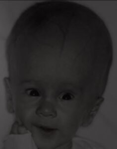
In the newborns, their ventricles dilate and enlarge the head circumference but in the case of adults, the ventricle enlarges, however, there is no increase in head circumference.
Physical signs and symptoms
- Nausea and Vomiting
- Sleepiness
- Irritability, loss of coordination or balance
- Poor feeding and lethargy
- Seizures and headache
- Blurred or double vision or Sunsetting of the eyes (Eyes fixed downward)
- Deficits in muscle tone and strength
- Loss of tactile response
- Poor growth and development
- Loss of memory power, concentration, and thinking skills
Parinaud syndrome: It is hydrocephalus and paralysis of upward gaze.
Complications
- If the condition is well advanced at birth, majorly brain damage & physical disabilities of the humans are likely.
- Other common complications include:
- Impairment Intellectual
- Neurological damage, like decreased function, movement, or sensation
- Problems with the artificial CSF drainage channel (such as surgical shunt), such as a kinking or blockage of the shunt tubing
- Infection at the site of the shunt occurs
Arnold-Chiari malformation: It is defined as the caudal extension of the medulla and cerebellar vermis through the foramen magnum. It a cause of hydrocephalus mainly non-communicating type.
Dandy-Walker malformation: It is defined as the partial or complete absence of the cerebellar vermis. Cystic dilation of the fourth ventricle. It is the cause of hydrocephalus, mainly non-communicating hydrocephalus.
When to visit a doctor or consultant
There are a few common signs & symptoms that arise and goes to a consultant or doctor
- High pitched cry
- Problem with feeding or suckling
- Breathing problems
- Seizures and unexplained, recurrent vomiting
Risk factors
- Newborns
- In congenital hydrocephalus or shortly after birth may occur because of any of the following risk factors, such as
- Bleeding disorder within the ventricles in your heart, a common possible complication of a premature birth baby.
- Newborns babies have abnormal development of the central nervous system (CNS) that may cause obstruct the flow of cerebrospinal fluid (CSF)
- During the pregnancy period of infection in the uterus occurs, like rubella or syphilis, may cause inflammation in fetal tissues in the brain
- In congenital hydrocephalus or shortly after birth may occur because of any of the following risk factors, such as
- Other factors
- Other factors that can role to develop hydrocephalus among any age group include:
- Tumors or lesions occur in the brain or spinal cord
- Central nervous system (CNS) infections, such as bacterial meningitis or mumps, etc.
- Bleeding disorder in the brain from a stroke or head injury cases
- Other traumatic injuries to the brain
- Other factors that can role to develop hydrocephalus among any age group include:
Diagnosis of the hydrocephalus
It is usually based on the
- History taking (signs and symptoms)
- General physical examination test
- Neurological examination test
- Brain imaging tests (CT-Scan or MRI or Ultrasound)
Treatment
There are two common surgical treatments are following
- Endoscopy of the third ventriculostomy
- Shunt
- Complications surgery
- In this condition, several complications may arise, so it is a suitable method for the treatment of complications
- Other treatments: In this case, a specific condition required a specific consultant or doctor, or teacher that may help to treatment of hydrocephalus such as
- Occupational therapist
- Pediatrician or psychiatrist
- Pediatric neurologist
- Developmental therapist
- Mental health provider
- Social worker
- Special education teacher