What is the inguinal canal?
The inguinal canal is an oblique placed inter-muscular passage that is around 4 cm long lying above the medial half of the inguinal ligament.
The extension of the inguinal canal from the deep inguinal ring to the superficial inguinal ring. It is directed downward, forward, and medially in the body.
Inguinal rings
There is 2 inguinal ring present, they are deep and superficial inguinal rings.
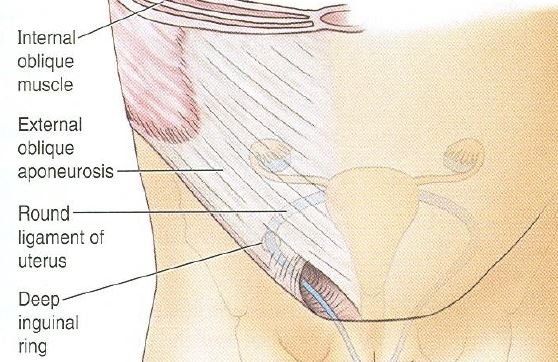
Deep inguinal ring
It is an oval-shaped opening in the fascia transversalis and it lies around 1.25 cm above the mid inguinal point. From its margins, the fascia transversalis is extended into the canal like a sleeve, the internal spermatic fascia, throughout the structures that pass through the ring.
The structure passage throughout the deep inguinal rings is a spermatic cord in males, In the case of females, round ligament of the uterus obliterated remains of processus vaginalis, and lymphatics from the uterus.
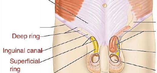
Superficial Inguinal Ring
It is a triangular space in the aponeurosis of the external oblique muscle and lies above and lateral to the pubic crest.
The pubic crest layout the base of the triangle and the side (lower/lateral margins and upper/medial) of a triangle which are called crura, which meet laterally to form an obtuse apex.
The structures passing throughout the inguinal ring are in males such as the spermatic cord and ilioinguinal nerve, and in the case of females, there are round ligaments of the uterus and ilioinguinal nerve.
Boundaries of the inguinal canal
- Anterior wall (from superficial to deep):
- Skin (whole extent)
- Superficial fascia in the whole extent (whole extent).
- External oblique aponeurosis (whole extent).
- Internal oblique muscle fibers (lateral one-third).
- Posterior wall (from deep to superficial):
- Fascia transversalis (whole extent).
- Conjoint tendon (medial two-third).
- Reflected part of the inguinal ligament (medial-most part).
- Roof:
- lower arched fibers of transversus abdominis muscles and internal oblique muscles.
- Floor:
- To a whole extent a grooved upper surface of the inguinal ligament.
- It is bounded by the abdominal surface of the lacunar ligament which is the medial end.
Contents in the inguinal canal
- In males: Spermatic cord and ilioinguinal nerve.
- In females: Round ligament of the uterus and ilioinguinal nerve.
Spermatic cord
The spermatic cord is a collection of structures and that course to and fro from the testis through the inguinal canal.
It is extending from the deep inguinal ring to the posterior border of the testis of the male. It is covered by 3 fascial layers, they are including; internal spermatic fascia, cremasteric fascia, and external spermatic fascia.
Constituents in the spermatic cord
- The spermatic cord consists of various structures, including;
- Ductus deferens (posterior part).
- Arteries:
- Testicular artery (from the abdominal aorta).
- Cremasteric artery (from an inferior epigastric artery).
- Artery to ductus deferens (from an inferior vesical artery).
- Veins: the pampiniform venous plexus.
- Lymphatics ( pre-and para-aortic nodes and some external iliac nodes).
- Nerves:
- Genital branch of the genitofemoral nerve.
- Sympathetic fibers
- Remains of processus vaginalis.
Mechanism to maintain the integrity of the inguinal canal
A site of potential frailty in the lower part of the anterior abdominal wall. The herniation of the abdominal viscera. Normally prevented by the following mechanism:
- Flap-valve Mechanism
- Guarding of the Inguinal Rings
- Shutter Mechanism
- Slit-valve Mechanism
- Ball-valve Mechanism
Inguinal triangle (Hesselbach’s triangle)
The inguinal triangle is located deep to the posterior wall of the inguinal canal of the body. It is noticed on the inner aspect of the lower part of the anterior abdominal wall.
Boundaries of the inguinal triangle
- Medial: Lower about 5 cm of the lateral border of the rectus abdominis muscle.
- Lateral: Inferior epigastric artery.
- Inferior: Medial half of the inguinal ligament.
- Floor: It is covered by the peritoneum, extraperitoneal tissue, and fascia transversalis.
Applied anatomy
- Inguinal hernias
- Indirect inguinal hernia (congenital and acquired)
- Sac enters through the deep inguinal ring lateral to the inferior epigastric vessels
- Direct inguinal canal hernia (medial and lateral)
- Through the posterior wall of the medial inguinal canal to the inferior epigastric vessels
- Indirect inguinal hernia (congenital and acquired)
Coverings of the direct and indirect hernias
| Direct inguinal hernia | Indirect inguinal hernia |
| Extraperitoneal tissue | Extraperitoneal tissue |
| Fascia transversalis | Internal spermatic fascia |
| Conjoint tendon (in medial direct hernia) | Cremasteric fascia |
| Cremaster fascia (in lateral direct hernia) | External spermatic fascia |
| External spermatic fascia | Skin |
| Skin |
[embeddoc url=”https://notesmed.com/wp-content/uploads/2021/05/Inguinal-canal.pdf” download=”all” cache=”off” text=”Complete pdf file-Download”]