What is the thyroid gland?
The thyroid gland is the largest endocrine gland and a weight of about 20 to 30 grams and increases during periods of pregnancy and menstruation.
The location of the thyroid gland is at the neck region; each lobe lying on either side of the lower part of the larynx and upper part of the trachea which is just opposite to the fifth cervical (C5) to the first thoracic (T1) vertebrae.
Capsules of the thyroid gland
- Thin fibrous (true) capsule: It is sent septa into the gland.
- False capsule: It is external to the true capsule in the glands.
-
- Splitting of pre-tracheal fascia in the body.
- It is attached to the hyoid bone in the midline & oblique line of thyroid lamina.
- The thickened false capsule is attached to the cricoid cartilage is called the ligament of Berry.
- In between 2 capsules, there are the following structures present such as thyroid vessels and parathyroid glands.
Presenting parts
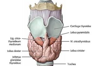
This gland is H-shaped or butterfly-shaped gland and consists of the following parts;
- Two lateral lobes
- An isthmus: It is connecting two lobes.
- Pyramidal lobe: It often presents and projects upward from the isthmus generally to the left of the midline.
- levator glandulae thyroideae: It is a fibrous or fibromuscular band that connecting the pyramidal lobe to the hyoid bone in the body.
Lateral lobes
It is abruptly pyramidal in shape with base, apex, three surfaces such as anterolateral, postero-lateral, and medial surface, and two borders include anterior and posterior borders.
Apex
- It is an upwards to the oblique line of the thyroid cartilage in the body.
- It is present between inferior constrictor muscle and sternothyroid muscle.
- Near the apex area, the superior thyroid artery, and external laryngeal nerve devide.
- The ligation of the artery close to the thyroid gland in the body.
Base:
- It extends or elongates up to the 5th to 6th tracheal ring in the body.
- It is related to the loop of the inferior thyroid artery & recurrent laryngeal nerve in the body.
- The ligation of the artery away from the thyroid glands.
Relations of the lobes of the thyroid gland
Anterolaterally
- Sternothyroid.
- Superior belly of omohyoid.
- Sternohyoid, and
- Anterior border of sternocleidomastoid muscles (SCM).
Posterolaterally
- Carotid sheath with the common carotid artery in the body
- Internal jugular vein
- The vagus nerve
Medially
- Larynx
- Trachea
- Pharynx
- The esophagus
- Cricothyroid muscle
- Inferior constrictor muscle
- External laryngeal nerve and a recurrent laryngeal nerve in the groove between the esophagus and trachea in the body.
- Cricoid cartilage and thyroid cartilage
The posterior border is related to the following structures;
- Medial & postero-lateral surface in the body.
- Superior & inferior parathyroid glands in the body.
- Anastomosis occurs in between the superior and inferior thyroid arteries.
Anterior border
- Antero-lateral surface and medial surface in the gland.
- It is related to the anterior descending branch of the superior thyroid artery in the body.
Relations of the Isthmus
The anterior surface of the isthmus
- Sternothyroids muscle
- Sternohyoids muscle
- Anterior jugular veins
- Fascia and skin
The posterior surface of the isthmus
- The second, third, and 4th tracheal rings in the body.
The upper border of the isthmus
- Arterial anastomoses occur between two superior thyroid arteries in the isthmus.
Lower border
- Inferior thyroid veins come out and thyroid ima artery (when present) enters through this lower border.
Blood supply
Arteries: There following artery supply the blood to the glands;
- Superior thyroid artery
- Inferior thyroid artery.
- Thyroid ima artery (sometimes).
- Accessory thyroid arteries:
- It is profuse anastomosis with one another on the surface of the thyroid gland in human beings.
- Superior thyroid artery:
-
- It is a branch of the external carotid artery and it descends downwards and forwards to the superior pole of the lobe, go along with an external laryngeal nerve.
- It supplies the upper 1/3rd of the lobe & upper half of the isthmus in the human.
- Inferior thyroid artery:
-
- It is a branch of the thyrocervical trunk in the neck region and it ascends behind the thyroid gland up to the cricoid cartilage.
- It turns medially and downwards to the posterior border of this gland.
- The recurrent laryngeal nerve crosses either in front of or beyond the artery or may pass between the branches and that gives 4 or 5 branches.
- It supplies the lower 2/3rd of the lobe and the lower half of the isthmus in the body.
- Thyroid ima artery:
-
- If present appears from the brachiocephalic trunk or the arch of the aorta in the body.
- It enters into the isthmus from below the area.
- Accessory thyroid arteries:
-
- It arises from tracheal and esophageal arteries in the body.
Venous drainage
- The superior thyroid vein – into the internal jugular vein.
- The middle thyroid vein – the internal jugular vein.
- Inferior thyroid vein-from inferior pole and the isthmus of the gland.
- Inferior thyroid veins anastomose occurs with one another as they descend in front of the trachea in the neck.
- left brachiocephalic vein drainage in the thorax.
Lymphatic drainage
- It is mainly into the deep cervical nodes in the neck region
- It also to the paratracheal lymph nodes
[embeddoc url=”https://notesmed.com/wp-content/uploads/2020/09/Thyroid-and-parathyroid.pdf” download=”all”]