What is the trachea?
The trachea is a fibrocartilaginous tube that is a passage of air. The trachea goes down (descends) as a continuation of the larynx and ends by dividing it into two principal bronchi.
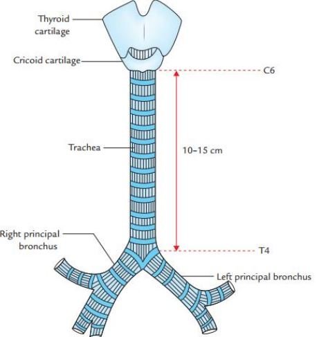
The trachea extent from the sixth cervical (C6) to the fourth thoracic (T4) in cadaver and supine position & the sixth cervical (C6) to the sixth thoracic (T6) in living individuals in standing position, and C6 to T3 vertebral level in newborns.

Dimensions
- Length – 10-15 cm.
- Breadth (External)
- In adult male 2 cm.
- In adult females 1.5 cm.
- Breadth (Internal):
- 12 mm in adult.
- The lumen of the trachea:
- The lumen of the trachea is larger in the cadavers than in living human beings.
- In newborn: 3mm at the third year of life and then increases lumen size by 1mm up to 12 years and remains constant.
The course of the trachea
- The trachea is the continuation of the larynx in the neck region.
- Begins at the inferior border of the cricoid cartilage at the level of the sixth cervical (C6) vertebra, about 5 cm above the jugular notch.
- It enters into the thoracic inlet in the midline.
- Passes downwards and backward beyond the manubrium to terminate by bifurcating into two principal bronchi.
- A little to the right side at the lower border of the fourth thoracic (T4) vertebra corresponding to the sternal angle.
Structure of trachea
It is made by incomplete C-shaped cartilaginous ring structures and connected by a strong fibro-elastic membrane.
The absence of cartilages on the posterior aspect permits the expansion of the esophagus during the deglutination process.
It contains 16-20 rings, 1st one is the broadest ring, and the last one is a triangular outline which is called carina. Involuntary muscles-trachealis present on the posterior surface of the trachea.
What is a carina?
It is a triangular process of the last ring of the trachea. Which hooks upward and surrounds the beginning of two principal bronchi and the carina is associated with cough reflex.
Relations In Neck (Cervical region)
In front:
- Skin
- Superficial and deep cervical fasciae in the neck
- Jugular venous arch crossing in the suprasternal space in the neck area.
- Overlapped by sternohyoid muscle & sternothyroid muscles.
- It is crossed by an isthmus of the thyroid gland in the neck area.
- Inferior thyroid veins and arteria thyroidea ima which is occasionally.
- The left brachiocephalic vein in children may arise in the neck region.
- The thymus gland in children
- Brachiocephalic artery present sometimes in children.
Behind:
- Oesophagus
- The recurrent laryngeal nerve on each side.
On each side, it is related to the following structures:
- A lobe of the thyroid gland is elongating up to the 5th or 6th tracheal ring.
- The common carotid artery present in the carotid sheath.
Relation In Thorax
In front:
- Manubrium sterni.
- Pre-tracheal fascia containing inferior thyroid veins in the thoracic region.
- It is crossed by left brachiocephalic vein.
- Superior vena cava.
- Deep cardiac plexus.
- Aortic arch
- Brachiocephalic, and left common carotid artery.
Behind:
- Oesophagus.
- Left recurrent laryngeal nerve (tracheo-oesophageal groove).
- Vertebral column.
On the right side:
- Right lung
- Mediastinal pleura.
- Right vagus nerve.
- Arch of the azygos vein.
On the left side:
- The arch of the aorta.
- left common carotid artery
- Left subclavian arteries.
- Left lung.
- Left vagus and phrenic nerves.
Arterial supply
- Inferior thyroid artery
- Bronchial arteries.
Venous drainage:
- Inferior thyroid vein.
Lymphatic drainage:
- Pre and paratracheal lymph node.
Nerve supply:
- Parasympathetic: The vagus and recurrent laryngeal nerve.
- Sympathetic: Upper 4 or 5 thoracic segments of the spinal cord.
[embeddoc url=”https://notesmed.com/wp-content/uploads/2020/10/Trachea-and-Broncial-tree.pdf” download=”all”]