Shoulder joint
The shoulder joint is a complex joint consist of the following four basic articulations, they are shown below,
- Glenohumeral joint
- Acromioclavicular joint
- Sternoclavicular joint
- Scapulothoracic articulation/ scapulothoracic linkage (which is the functional linkage between the scapula and thorax).
The impairment of any one of these joints leads to functional defects of the whole complex joint in your body.
SHOULDER JOINT (GLENOHUMERAL JOINT)
The shoulder joint is present between the head of the humerus bone and the glenoid cavity of the scapular bone. It is the most movable joint in our human body and consequently one of the least stable & the most common joint to dislocate and to undergo recurrent dislocations of the joint in the human body. It is a ball-and-socket variety of synovial joints in the human body.

Articular surfaces
The shoulder joint is formed by the articulation of an enlarged round head of the humerus with the comparatively shallow glenoid cavity of the scapula. The glenoid cavity is deepened slightly but successfully or effectively by the fibrocartilaginous ring which is called the glenoid labrum.
Ligaments
There are following ligaments of the shoulder joint are given below;
- Capsular ligament (joint capsule).
- Glenohumeral ligaments.
- Coracohumeral ligament.
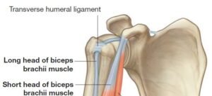
- Transverse humeral ligament.
- Accessory ligaments:
- Coracoacromial ligament.
- Coracoacromial arch.
Bursae related to the joint
Several bursae are related to this shoulder joint but important ones are the following below;
- Subscapular bursa.
- Subacromial bursa.
- Infraspinatus bursa.
Relations of the shoulder joint
Superiorly:
- Coracoacromial arch.
- Subacromial bursa.
- Supraspinatus muscle
- Deltoid muscle.
Inferiorly:
- The long head of triceps.
- Axillary nerve
- Posterior circumflex humeral vessels.
Anteriorly:
- Subscapularis.
- Subscapular bursa
- Coracobrachialis.
- The short head of biceps brachii
- Deltoid muscle
Posteriorly:
- Infraspinatus.
- Teres minor.
- Deltoid muscle.
Arterial supply
- Anterior and posterior circumflex humeral arteries.
- Suprascapular artery.
- Subscapular artery.
Nerve supply
- Axillary nerve.
- Suprascapular nerve.
- Musculocutaneous nerve.
Factors responsible for stability to the joint
- Rotator cuff (musculotendinous cuff).
- Coracoacromial arch.
- The long head of the biceps tendon.
- Glenoid labrum.
Movements of the shoulder joint
It has more freedom of mobility or flexibility than any other type of joint in the body, due to the following factors:
- Laxity of the joint capsule.
- Articulation between the comparatively large humeral head and smaller and shallow glenoid cavity.
- Flexion and extension.
- Abduction and adduction.
- Medial and lateral rotation.
- Circumduction.
- The flexion and extension or hyperextension occur in the sagittal plane nearby the frontal axis.
- The abduction & adduction appear in the frontal plane around the sagittal axis.
- The medial rotation & lateral rotation appears in the transverse plane around the vertical axis.
- The circumduction is really only a combination of all the above mentions movements.
Applied anatomy
Dislocation of the shoulder joint:
- It mostly occurs inferiorly due to the fact that the joint is least supported on this aspect.
- Injures the axillary nerve due to the fact that its close relation to the inferior part of the joint capsule.
Frozen shoulder (adhesive capsulitis):
Rotator cuff disorders:
[embeddoc url=”https://notesmed.com/wp-content/uploads/2020/08/Shoulder-Joint-Complex.pdf” download=”all”]