What is the pons?
The pons is a large bulky transverse mass of the brainstem present between the midbrain and medulla oblongata.
On either side, the pons is continual to the middle cerebellar peduncle, thus create a bridge between the two cerebellar hemispheres, hence its name pons (L. pons = bridge).
External features
The ventral and dorsal surfaces present in the pons and have two borders; superior and inferior borders.
Ventral surface of pons
The convex ventral surface in both directions, i.e., from before backward and from side to side. It is striated transversely because of underlying pontocerebellar fibers.
In the median plane, it presents a vertical (upright) groove, the basilar groove which lodges the basilar artery.
The trigeminal nerve is connected to this surface area by two roots including, a small motor and a large sensory root (the motor root lies medial to the sensory root).
Rostrally, Midbrain and pons formed a junction which is marked by cerebral peduncles and the intervening interpeduncular fossa; caudally the junction of the pontomedullary is marked by a shallow groove.
In this groove, from medial to lateral, the abducent (VI) nerve, facial (VII) nerve, and vestibulocochlear (VIII) nerves emerge.
The superior cerebellar arteries turn along the superior border, intervening in the middle of the oculomotor and trochlear nerves. The anterior inferior cerebellar arteries turn around the inferior (lower) border.
Dorsal surface of pons
It is surrounded by the cerebellum and divided from it by the cavity of the 4th ventricle. It is triangular in shape and forms the upper part of the floor of the fourth (4th) ventricle in the brain.

Internal features of the pons
The ventral part or basilar part is continuous inferiorly with the pyramids of the medulla oblongata & on each side with the middle cerebellar peduncle which connects the cerebellum.
The dorsal part or tegmental part is a direct upward continuance of the medulla oblongata excluding the pyramids of the brain.
Basilar part
It is formulated of the fibers of longitudinal bundles, the transverse fibers, and the pontine nuclei:
- Longitudinal bundles of fibers include the following they are; corticopontine, corticonuclear, and corticospinal fibers.
- The corticopontine fibers pass on in the ipsilateral pontine nuclei.
- The fibers of corticonuclear terminate in the contralateral (and to some extent ipsilateral) motor nuclei of the cranial nerves in the pons.
- The fibers of the corticospinal connect toward the lower (inferior) part of the pons and form the pyramids of the medulla.
- The fibers of the transverse arise in the pontine nuclei and cross across from the side to form the middle cerebellar peduncle -pontocerebellar fibers.
- Pontine nuclei are scattered out of the longitudinal and transverse fibers.
Tegmental part
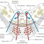
It is traversed by a number of ascending tracts and descending tracts and contains a decussation of transversely running fibers, which is called the trapezoid body.
It also contains the number of nuclei such as nuclei of trigeminal (V), abducent (VI), facial (VII), and vestibulocochlear (VIII) nerves.
Since the structure of the tegmentum differs in the inferior (caudal) and superior (cranial) parts of the pons of the brainstem, it is studied by examining transverse sections at these two levels in the pons.

Arterial supply
- Many (pontine) branches from the basilar artery.
- Anterior inferior cerebellar artery.
Applied Anatomy of pons
Millard-Gubler syndrome (Medial inferior pontine syndrome)
- It due to the fact that a lesion in the lower (inferior) part of the pons, that includes the pyramidal tract, the appearing fibers of the abducent nerve, and facial nerves. Ipsilateral medial squint (inward diversion of an eye towards the side of the lesion) due to abducent nerve involvement.
- Ipsilateral facial palsy, due to participation of facial nerve fibers. The contralateral hemiplegia, due to the fact that involvement of the corticospinal tract in the brain.
Pontocerebellar angle syndrome
- In this syndrome, there are present following symptoms such as tinnitus, progressive deafness, and vertigo due to damage of the VIII cranial nerve.
- Ipsilateral ataxia and staggering gate due to the fact that compression of the cerebellar peduncle in the brain.
- Ipsilateral lower motor neuron (LMN) type of facial palsy, due to the participation of facial nerve.
- Ipsilateral loss of pain and temperature perception and loss of corneal reflex due to the perception of the spinal tract and nucleus of the trigeminal nerve in the brain
[embeddoc url=”https://notesmed.com/wp-content/uploads/2020/11/Pons.pdf” download=”all”]