What is the pituitary gland?
The pituitary gland is the most complex endocrine gland. It is also called the master gland.
Location of the pituitary gland
It is a small endocrine gland situated in the hypophyseal fossa on the superior surface of the body of the sphenoid bone (sella turcica). The gland is suspended from the floor of the third ventricle of the brain by an Infundibulum(narrow stalk).
Shape and Measurement
A gland is Oval or pea in shape and its length (anteroposteriorly): 8 mm and breadth (Transversely): 12 mm.
Weight: About 500 mg.
Parts of Pituitary Gland
The gland consists of two distinct parts, they are:
- Adenohypophysis (anterior lobe)
- Neurohypophysis (posterior lobe)
These two parts anterior and posterior lobe differ from each other embryologically, morphologically, and functionally.
Development
Adenohypophysis (anterior lobe):
The adenohypophysis develops as a diverticulum (Rathke’s pouch) from the ectodermal lining of the stomodeum. Due to the rapid growth of the sphenoid, the communication with the roof of the pharynx disappeared. The uppermost part of the original diverticulum remains as a cleft which separates the pars anterior and pars intermedia.
Neurohypophysis (posterior lobe):
The neurohypophysis (posterior part) develops as downward growth from the floor of the diencephalon.
Adenohypophysis consists of:
- Pars anterior (pars distalis)
- Pars intermedia, and
- Pars tuberalis
Neurohypophysis consists of:
- Pars posterior, and
- Infundibulum
Relations
- Superior (Above): Diaphragma sellae, optic chiasma, Infundibular recess of the third ventricle.
- Inferior (Below): Hypophyseal foss and Sphenoidal air sinuses.
- Lateral (On each side): Cavernous sinus and its contents.

Blood supply
Arterial supply: The arterial supply by branches of the internal carotid artery such as;
- Superior hypophyseal artery, one on each side of the gland.
- Inferior hypophyseal artery, one on each side of the gland.
The arterial supply of the pars anterior that exhibits hypothalamohypophyseal portal circulation.
Venous drainage:
Short veins from the pituitary gland drain into neighboring dural venous sinuses such as cavernous and inter-cavernous sinuses.
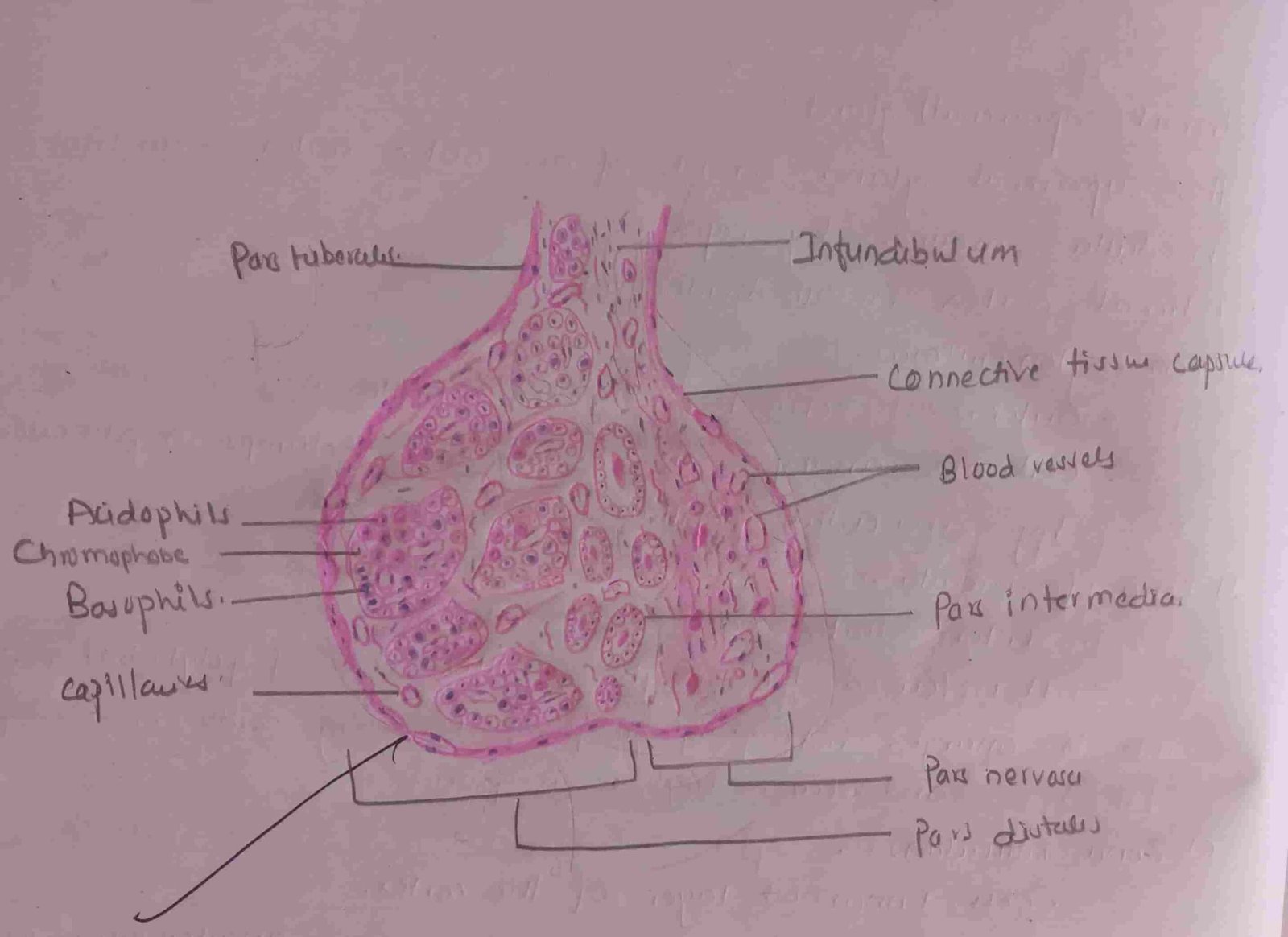
Micro anatomy
- Adenohypophysis consists of:
- Pars anterior (pars distalis)
- Pars intermedia, and
- Pars tuberalis
- Pars anterior (pars distalis):
- It is a major part of the adenohypophysis (anterior lobe) and consists of an anastomosing cord of the cells that are surrounded by sinusoids. The cells forming cords are shown below,
- Chromophobes
- Chromophils
- It is a major part of the adenohypophysis (anterior lobe) and consists of an anastomosing cord of the cells that are surrounded by sinusoids. The cells forming cords are shown below,
- Chromophobes:
- They are smaller in size and have little affinity for histological stain, so it is called chromophobe. These cells contain little cytoplasm which usually does not have any granules. These chromophobes are considered exhausted cells (or inactive phase of chromophils).
- Chromophils:
- More stainable and larger than chromophobes, and possess secretory granules. They are two types of names, acidophils, and basophils.
- Acidophils: On the basis of function, the acidophils are classified into the following types:
- Somatotrophs: They regulate body growth by secreting growth hormones (GH).
- Corticotrophs: They secreting ACTH (Adrenocorticotrophic hormone) and act on the suprarenal gland.
- Mammotrophs: Secreting prolactin hormone and act on the mammary gland.
- Basophils: on the basis of the function, they are classified into,
- Thyrotrophs: that secrets TSH (Thyroid-stimulating hormone) and act on the thyroid gland.
- Gonadotrophs: Acts on the gonads by secreting the following hormone:
- FSH (Follicle-stimulating hormone)
- LH (luteinizing hormone) in females
- ICSH (interstitial cell-stimulating hormone) in the case of a male.
- Pars intermedia:
- It is a narrow zone and consisting of small cells. It is separated from pars anterior by inter-glandular cleft. The cells of the pars intermedia that secrets MSH (Melanocyte-stimulating hormone).
- Pars tuberalis:
- Upward tubular extension of the anterior surrounding part of the infundibulum and the cells within it are called basophilic, which contain some granules. The function is uncertain.
- Neurohypophysis:
- It does not contain neurons but develops from a ventral outgrowth of the floor of the diencephalon. It consists of a narrow infundibulum (neural stalk) and swollen pars nervosa (pars posterior or infundibular process).
- Pars nervosa:
- It consists of pituicytes and non-myelinated nerve fibers that originate from the cell bodies of the neurons located in the hypothalamus. The nerve fibers constitute the hypothalamohypophyseal tract. It passes to the pars nervosa and terminates with swollen ending close to the blood capillaries. It does not secrets any hormone but simply serves as a depot for storage of the hormones (antidiuretic and oxytocin hormone) which are secreted by the hypothalamus.