What is the pharynx?
The pharynx is a funnel-shaped fibromuscular tube structure that is lined by a mucous membrane. It extends from the base of the skull to the sixth cervical (C6) vertebra of the body.
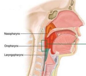
The location of the pharynx is behind the nose which is called the nasopharynx, behind the mouth is known as the oropharynx, and behind the larynx is known as the laryngopharynx.

Boundaries of Pharynx
- Roof: It is formed by the body of the sphenoid and basilar part of the occipital bone in the human.
- Inferiorly: It is continuous with the esophagus just opposite to the sixth cervical (C6) vertebra.
- Posteriorly: It is supported by the upper six cervicals (C1-C6) vertebra, prevertebral muscles, & fascia.
- Anteriorly: It communicates with the oral cavity, nasal cavity, and larynx in human beings.
- On each side: It is attached or connected to the medial pterygoid plate, pterygomandibular raphe of the mandible, tongue, hyoid bone, thyroid, and cricoid cartilage in the body.
The pharynx is related to the styloid process, internal carotid arteries, common carotid artery, & external carotid arteries. The pharynx which it communicates with the middle ear cavity with a eustachian tube (auditory tube ).
Sub-division
- Nasopharynx
- Oropharynx
- Laryngopharynx
Nasopharynx
It is present just behind the nasal cavity which extends from the base of the skull superiorly to the soft palate inferiorly. It communicates anteriorly with the nasal cavity in the body.
- The roof and posterior wall form a continuous surface & present the following structures:
- Pharyngeal or nasopharyngeal tonsil.
- Pharyngeal bursa.
- Lateral wall
- Pharyngeal opening of the eustachian tube (auditory tube) about 1.25 cm beyond and slightly below the level of the inferior nasal concha.
- Tubal elevation- It guards the upper and posterior margin of the eustachian tube (auditory tube) and which overlies the tubal tonsil.
- Two mucous folds pass downward, they are the following;
- Salpingopharngeal fold.
- Salpingopalatine fold (formed by the Levator veli palatini muscle)
- Pharyngeal recess (fossa of Rosenmuller): The mucous covered deep depression beyond the tubal elevation.
- Floor: It communicates with the oropharynx via. the pharyngeal isthmus.
- Boundaries of pharyngeal isthmus:
- Anterior: The posterior surface of the free border of the soft palate.
- Posterior: Mucous elevation which is formed by palatopharyngeal sphincter (passavant’s ridge).
- On each side: Palatopharyngeal arch in the isthmus.
- Boundaries of pharyngeal isthmus:
Oropharynx
It is present beyond the oral cavity and in front of the 2nd & 3rd Cervical vertebra. It extends from the superiorly soft palate to the tip of the epiglottis inferiorly.
At below, it communicates with the laryngopharynx which is a subdivision of the pharynx. It communicates anteriorly with the oral cavity via. the oropharyngeal isthmus.
Laryngopharynx
It is the lower part of the pharynx situated beyond the larynx. It elongates from the epiglottis (upper border) to the cricoid cartilage (lower border).
It anteriorly communicates with the laryngeal cavity via. the laryngeal inlet and inferiorly communicates with the esophagus. The piriform fossa present on each side.
Boundaries of laryngopharynx
- Anterior wall: The inlet of the larynx, & posterior surface of the cricoid cartilage & arytenoid cartilage
- Posterior wall: It is mainly by the fourth (C4) and sixth (C6) cervical vertebra
- Lateral wall: Piriform fossa
Structure of pharynx
From within outwards, they are the following;
- Mucosa:
- Nasopharynx: Pseudostratified ciliated columnar epithelium tissue
- Oro- and laryngopharynx: Stratified squamous nonkeratinized epithelium tissue
- Pharyngo-basilar fascia: It is formed fibrous sheet internal to the pharyngeal muscles
- Muscular coat
- Buccopharngeal fascia
Muscles of the pharynx
It consists of striated muscles which are organized in the outer circular and inner longitudinal layer.
The circular layer includes the superior constrictor muscle, middle constrictor muscle, and inferior constrictor muscle.
These muscles joined together posteriorly to form the pharyngeal raphe. It is anteriorly attached to the bones & ligaments which is related to the lateral margins of the nasal and oral cavities and the larynx of the body.
The longitudinal layer comprises the stylopharyngeus muscle, palatopharyngeus muscle, and salpingopharyngeus muscle.
Nerve supply of the pharyngeal muscles
Motor nerve supply
- All the muscles present in the pharynx are supplied by the cranial part of the accessory nerve through the pharyngeal plexus except for the stylopharyngeus muscle in the longitudinal layer.
- The longitudinal layer muscle called the stylopharyngeus muscle is supplied by the glossopharyngeal nerve.
- The inferior constrictor muscle also receives additional supply from the external laryngeal nerve and recurrent laryngeal nerve.
Sensory nerve supply
- Naso-pharynx: It is supplied by the maxillary nerve via. the pharyngeal branch of the pterygopalatine ganglion.
- Oro-pharynx: It is supplied by the glossopharyngeal nerve.
- Laryngopharynx: It is supplied by the internal laryngeal nerve.
Blood supply
Arterial supply
- Ascending pharyngeal, Ascending palatine, and tonsillar artery are branches of the facial artery.
- Greater palatine artery, pharyngeal and pterygoid branches of the maxillary artery, and Dorsal lingual branches of the lingual artery.
Venous supply
The vein of the pharynx forms a pharyngeal venous plexus which joins with the pterygoid venous plexus and which drains into the internal jugular vein.
[embeddoc url=”https://notesmed.com/wp-content/uploads/2020/09/Pharynx.pdf” download=”all”]