What is a Neuromuscular junction?
Skeletal muscles are innervated by motor nerves. The junction between a motor neuron and a muscle fiber is known as the neuromuscular junction (NMJ).
Or
Each branch of a motor nerve fiber terminates nearby the center of the muscle fiber to form a rounded structure known as a neuromuscular junction (NMJ) or myoneural junction or motor endplate.
Parts of Neuromuscular junction
- Presynaptic
- Synaptic cleft
- Postsynaptic
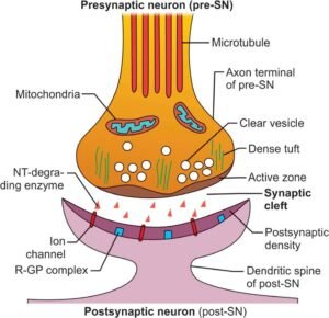

Presynaptic Portion (Axon Terminal):
Features of the presynaptic portion of NMJ are:
- As the axon of a motor neuron approaches or approximate the muscle fiber, it loses its myelin sheath and divides extensively into numerous fine branches of about 2 μm in diameter, called axon terminals.
- The axon terminals are surrounded by Schwann cells, known as teloglia (glial cells at terminals).
- Each terminal forms a junction with a single skeletal muscle fiber, midway along its length.
- Thus, each muscle fiber is supplied by single motor neuron terminal.
- The motor neuron, including its axon and axon terminals and the muscle fibers supplied by it, is called a motor unit.
- Each terminal is expanded at its end to form a knobby structure, known as a synaptic knob (terminal button).
- The synaptic knob (terminal button) contains plenty of mitochondria and neurotransmitter vesicles.
- The terminal button situated in the groove (synaptic trough) in the surface area of the muscle fiber, but outside the muscle cell membrane.
- The presence of a large or huge number of mitochondria indicates higher metabolic activity in the terminal region.
- The vesicles are clustered around specific points known as active zones.
- The membrane at the active zones is modified to form a dense bar that contains numerous voltage-gated Ca++ channels (mediate ACh release).
Synaptic Cleft in Neuromuscular junction:
- The gap in the middle of the terminal button and the muscle fiber, which is about 40–100 nm wide.
- The muscle fiber is covered by a layer of amorphous connective tissue known as the basement membrane or basal lamina.
- Consisting of collagen, glycoproteins, and other extracellular matrix proteins.
- The basement membrane in the cleft contains the enzyme acetylcholine-esterase (AChE), which is anchored to the collagen fibrils and is secreted into the basement membrane by the presynaptic terminal and the muscle fiber.
- AChE rapidly hydrolyzes ACh into acetate and choline.
- The basal lamina helps organize the presynaptic buttons with the postsynaptic junctional folds.
Postsynaptic Portion (End Plate Membrane):
- The sarcolemma lies directly under the terminal button.
- The area of the endplate membrane increases many times as it is thrown into several folds called junctional folds.
- The endplate membrane contains numerous Ach receptors (AchR), which are concentrated at the crests of the junctional folds.
- It also contains voltage-gated Na+ channels.
Neuromuscular transmission
- The arrival of an Action potential at the axon terminal of NMJ.
- Depolarization of the membrane of the terminal buttons.
- Activation and Opening of voltage-gated Ca2+ channels, allowing Ca2+ ions to diffuse from synaptic space to the interior of the nerve terminal.
- Ca2+ ions attract Acetylcholine vesicles to fuse with the neural membranes and release their content via. exocytosis.
- Acetylcholine combine with the nicotinic acetylcholine receptor causes conformational changes to the acetylcholine gated ion channel.
- Na+ ion rushes to the interior of the cell membrane.
- An influx of Na+ ions leads to depolarization of postsynaptic membrane- endplate potential (EPP).
- When EPP reaches a threshold potential gives rise to an action potential.
- The spread of action potential to the T- tubules, which leads to the contraction of muscles.
Myasthenia Gravis
The inability of the neuromuscular junction to transmit enough signals from the nerve fibers to the muscle fibers. A lessen in the number of AChR presents on the motor endplate due to the production of circulating autoantibodies against these receptors.
Muscle fatigue, drooping of the eyelids, difficulty in swallowing or chewing, and breathing. In general, occurs 1 in 20,000 people women are mainly affected. Men are affected less than women in a ratio of 2:3.
It’s an autoimmune disease, antibodies against nicotinic acetylcholine receptors. The EPP that occurs in the muscle fibers is too weak to stimulate it. If the disease is intense, the patient dies of paralysis of respiratory muscles.
[embeddoc url=”https://notesmed.com/wp-content/uploads/2020/10/Neuromuscular-Transmission.pdf” download=”all”]