What is leprosy?
Leprosy is a chronic granulomatous disease of humans being mainly involving cooler parts of the body of skin and nerve and also capable of affecting any tissue or organs causing bony deformities and disfigurements in untreated cases which is caused by the intercellular bacterium is called Mycobacterium leprae. It is discovered by Hansen in 1873, hence it is also called Hansen’s disease.

The disease progresses as hypopigmented or erythematous skin patches with definite loss of sensation and primarily infects Schwann cells in the peripheral nerves leading to nerve damage and the development of deformities and disabilities. So, this is how the disease progresses.
The incubation period of leprosy patients has a long average of 3-5 years but varying between 2 and 40 years. The longer incubation period is due to the long generation time of the lepra bacilli. In the cases, the lepromatous cases have a prolonged incubation period than tuberculoid cases.
Classification of leprosy
Leprosy can be classified into various categories based on the bacteriological, clinical, immunological, and histological features of the patients.
- Madrid classification (1953)
- Lepromatous leprosy
- Tuberculoid leprosy
- Dimorphous leprosy
- Indeterminate leprosy
- Ridley–Jopling system of classification
- Tuberculoid polar Leprosy (TT)
- Borderline Tuberculoid (BT)
- Midborderline (BB)
- Borderline Lepromatous (BL)
- Lepromatous polar Leprosy (LL)
- WHO operational classification of leprosy
- Paucibacillary (TT, BT)
- Multibacillary {mid-borderline (BB), BL, LL}
Lepromatous leprosy
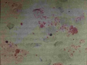
These cases are seen where host resistance is low to lepra bacilli. It is a multibacillary disease when a large number of low acid-fast bacilli are present in large clumps (globi) inside the foam macrophages (lepra cells) and few lymphocytes.
The clinical features; symmetrical with an irregular margin of the skin lesion, lesions appear as xanthomas like papules or plaques or superficial nodular lesions (lepromata). Thickening of the skin on the face produces leonine facies, loss of eyebrows/lashes. The lepromin test should be done then the test would be negative.
Tuberculoid leprosy
These cases are seen in patients with a high degree of resistance to lepra bacilli. It is a paucibacillary disease that has scanty bacilli in the lesions, organized granulomas, and multi-nucleated giant cells.
The skin lesions are few, asymmetric, and sharply demarcated, with macular anesthetic patches. In these cases, nerve involvement occurs earlier and nerves are often enlarged and thickened and nerve abscesses may be seen.
The ulnar nerve is the most commonly involved nerve and followed by the post-auricular nerve but the medial nerve is never involved. The lepromin test should be done test would be positive.
Other types of leprosy
- Borderline type of leprosy is seen in patients with characteristics between lepromatous and tuberculoid types. They may shift to either LL or TT type depending on the chemotherapy or alterations in resistance to the host.
- Indeterminate type of leprosy is seen those have early unstable cases with one or two hypopigmented macules & impairment of definite sensory. In this condition, the lesions are bacteriologically negative.
- A pure neuritic type of leprosy is seen in patients who develop neural involvement without any skin lesion in the body. This case is also bacteriologically negative.
- Midborderline type of leprosy shows multiple lesions around 10 to 30 and has variable sizes. In these cases, surface changes should be dull or slightly shiny. The hair growth should be diminished and the lepromin test should be done test would be negative.
Signs and symptoms
The signs and symptoms of specific leprosy explained above but common signs and symptoms are following below;
- Nodules on the skin
- Discolored patches of the skin
- Dry, thick, or stiff skin
- Painless ulcer on the soles of feet
- Painless swelling or lumps on the earlobes or faces
- Loss of eyelashes/eyebrow
Mode of transmission
- Multiple routes of transmission and portal of entry are either nose or skin.
- Nasal droplet infection
- Contact transmission (skin): direct and indirect contact.
- Direct dermal inoculation
Epidemiology
The highest new case detection rates are in India, Brazil, the Democratic Republic of Congo, Tanzania, Nepal, Madagascar, Angola, and the Central African Republic.
The South-East Asia Region accounts for 74% of new cases worldwide – the major burden being from India, Bangladesh, Indonesia, and Nepal. India and Nepal account for about 62% of the world’s leprosy.
Risk factors
- Genetic predisposition
- HLA-DR 2, 3 associated with tuberculoid form
- HLA-DQ 1 associated with lepromatous form
- Other factors
- Poverty
- Living in an endemic area
- Living in the same house with a leprosy patient
- Other diseases that compromise immune function
Immunology
- Innate immunity: shows a high degree of innate immunity to the lepra bacilli so that only a minority of these infected develop clinical disease.
- Humoral antibodies: It is produced against the various lepra antigen. In the case of M. leprae being intracellular which have a minor role in the disease control
- Cell-mediated Immunity: It is a vital role in the control of the disease condition.
- The tuberculoid disease is the result of high cell-mediated immunity with a largely Th1 type immune response.
- Lepromatous leprosy is characterized by low cell-mediated immunity and humoral TH2 response.
Complications
The complications of the patients are two types such as deformities and allergic response (lepra reactions).
- Deformities:
- It is about 25% of untreated cases develop deformities due to the course of time in which arise due to nerve injury leading to muscle weakness or paralysis, or disease process (eyebrow/lashes loss, facial deformities), or injury (ulcers) or infection.
- Some common deformities are; in the face (Leonine facies, sagging face, loss of eyebrow/lashes, saddle nose, and corneal ulcers), in the hand (wrist drop and claw hand), in feet (foot drop, inversion of the foot, clawing of toes and plantar ulcers).
- Lepra reactions:
- It is an immunological (allergic) type of acute exacerbation that occurs throughout its course so-called lepra reactions.
- In these reactions there are two types of reactions present such as reversal reactions (type 1 reaction) and erythema nodosum leprosum (type 2 reactions).
- Reversal reaction:
- It occurs in borderline leprosy and characterized by edema and erythema of existing skin lesions, the formation of new skin lesions, neuritis, and edema of the hands, feet, and face. The presence of inflammatory infiltrates with a predominance of CD4+ T cells.
- Treatment: Contact your doctor or consultant and patients usually respond well to glucocorticoids.
- Erythema nodosum leprosum(ENL):
- It occurs in lepromatous patients and characterized by Fever, chills, anorexia, malaise, subcutaneous tender nodules, lymphadenitis, arthritis, orchitis, iritis.
- In this case, the TH2 response is predominant with an increased level of IL-6 and IL-8. The tumor necrosis factor-α (TNF- α) plays a central role in the immunological reaction.
- Treatment: Started with glucocorticoids and thalidomide or clofazimine should be initiated for nonresponsive or recurrent conditions.
Management
- Combination chemotherapy: dapsone, rifampin, and clofazimine.
- Antiinflammatory for reactions: steroids, thalidomide
- Supportive care, eyes, hands, feet (to prevent tissue injury related to the neurologic deficit)
WHO recommended Multidrug therapy
The drugs should be used only after a doctor or consultant prescription.
- Paucibacillary case
- Rifampicin 600 mg/ month for 6 months
- Dapsone 100mg / day for 6 months
- For a patient with a single lesion a single dose of Rifampicin (600mg), Ofloxacin (400mg), and Minocycline (100mg)
- Multi bacillary case
- Rifampicin 600 mg/month for 2 or more years
- Dapsone 100 mg/day for 2 or more years
- Clofazimine 300 mg / month + 50 mg / day for 2 or more years.