What is the hip joint?
The hip joint is the largest ball and socket type of synovial joint between the head of the femur and the acetabulum of the pelvic bone. The functions of the hip joint are:
- To support the body weight during standing
- It helps to transmit the forces which are generated by movements of trunk femur during walking.

Articular surfaces
- Spherical head of the femur.
- The lunate side or surface of the acetabulum of the pelvic bone.
- The joint is a multi-axial ball and socket joint designed for stability and weight-bearing at the expense of mobility.
Ligaments
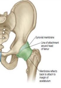
- Capsular ligament (joint capsule).
- Iliofemoral ligament (ligament of Bigelow).
- Pubofemoral ligament.
- Ischiofemoral ligament.
- Transverse acetabular ligament.
- Acetabular labrum.
- Ligamentum teres femoris (It is also called round ligament of the head of the femur).
Stability of the hip joint
- The depth of the acetabulum and narrowing of its mouth which is caused by the acetabular labrum.
- Three strong ligaments (iliofemoral, pubofemoral, and ischiofemoral) strengthening the capsule of the joint.
- Strength of the surrounding muscles, e.g., gluteus medius, gluteus minimus, etc.
- The length and inclination of the femoral neck.
Relations
Anteriorly:
- The tendon of the iliopsoas muscle is separated or differentiated from the joint by a synovial bursa, pectineus (lateral part), straight head of the rectus femoris.
- The femoral nerve in the groove between the iliacus and the psoas.
- Femoral artery in front of the psoas tendon.
- A femoral vein in front of the pectineus.
Superiorly:
- Reflected head of rectus femoris medially.
- Gluteus minimus, gluteus medius, and gluteus maximus laterally.
Posteriorly:
- Piriformis muscle, obturator externus muscle, superior and inferior gemelli, obturator internus muscle, quadratus femoris, and gluteus maximus muscle of this region.
- Superior gluteal nerve and vessels above the piriformis.
- Inferior gluteal nerve and vessels below the piriformis.
- The sciatic nerve, posterior cutaneous nerve of the thigh, and nerve to quadratus femoris.
Inferiorly:
- Pectineus.
- Obturator externus.
Bursae of the hip joint:
These are seven in number: four under gluteus maximus, one under gluteus medius, one under gluteus minimus, and one under psoas tendon as under:
- Between gluteus maximus and smooth area of the ilium lying between the posterior curved line and the outer lip of the iliac crest.
- Trochanteric bursa: Between gluteus maximus and lower part of the outer aspect of the greater trochanter.
- Ischial bursa: Between gluteus maximus and ischial tuberosity.
- Gluteofemoral bursa: Between the tendon of gluteus maximus and vastus lateralis.
- One bursa under the cover of gluteus medius between it and the upper part of the lateral aspect of the greater trochanter.
- One bursa under the gluteus minimus between it and the anterior aspect of the greater trochanter.
- The one between the iliac pubic bulge or eminence and the psoas major tendon. It is called subpsoas bursa. In 10% of individuals, the psoas bursa communicates with the synovial cavity of the hip joint through a gap in the thin part of the capsule between the iliofemoral and pubofemoral ligaments.
Movements
The hip joint is a multiaxial joint and permits the following types of movements are listed below:
- Flexion and extension.
- Abduction and adduction.
- Medial and lateral rotation.
- Circumduction (combination).
Applied anatomy
- Dislocation of the hip joint.
- Perthes’ disease (pseudocoxalgia).
- Osteoarthritis.
- Fractures of the neck of the femur.
[embeddoc url=”https://notesmed.com/wp-content/uploads/2020/09/Hip-Joint.pdf” download=”all” cache=”off”]