What is an eye?
The eye is an organ of vision(sight) and consists of the eyeball and the optic nerve. There are various components that include but are not so limited to the cornea, lens, retina, iris, pupil, optic nerve, choroid, and vitreous, etc. The cornea is transparent in the front part of the eye and that covers most of the part of the eye that focuses and transmits light into the eye.

Orbit
It contains the eyeball and its accessory visual structures (L., adnexa oculi). A pair of pyramidal-shaped bony cavities and located on either side of the root of the nose. The orbit provides sockets for rotatory movements of the eyeballs.
Boundaries of the orbit
Medial wall (thinnest):
- Frontal process of the maxilla.
- Lacrimal process of maxilla.
- An orbital plate of the ethmoid.
- Body of sphenoid.
Lateral wall (strongest):
- Orbital surface of the zygomatic bone in front.
- Orbital surface of the larger (greater) wing of sphenoid behind.
Floor:
- Orbital surface of the body of the maxilla.
- Orbital surface of the zygomatic bone, anterolaterally.
- Orbital process of the palatine bone, posteromedially.
Roof:
- An orbital plate of the frontal bone in front
- Lesser wing of the sphenoid behind.
Orbital Margins
- Supraorbital margin
- Infraorbital margin
- Medial orbital margin
- Lateral orbital margin
Orbital fascia or periorbita
The orbital fascia is a periosteum of the bony orbit. It is a line between the bony boundaries of the orbit and forms a funnel-shaped fascial sheath that encloses the orbit contents.
It is loosely attached to the bones, hence can be easily stripped off especially from the medial wall and roof of the orbit. At the optic canal and superior orbital fissure, it becomes continuous with the periosteum lining the interior of the skull (endocranium).
At the infraorbital fissure & orbital margins, it becomes continuous with the periosteum surrounding the external surface of the skull (periosteum).
Contents of the orbit of Eye
There are various components are present such as eyeball (most important content), Muscles, Fascia bulbi, Nerves (Optic, Oculomotor, Trochlear, Abducent, Ophthalmic, Ciliary ganglion), Ophthalmic artery, Ophthalmic veins, Lacrimal gland, Orbital fat.
The visual and orbital axis of the eye
- Visual axis: The line passes through the center of the anterior and posterior poles of the eyeball in the body.
- Orbital axis: The line passes through the optic canal and the center of the base of the orbit. Visual and orbital axis makes an angle of 20-300 with each other.
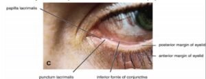
Lacrimal apparatus
The lacrimal apparatus concerned with secretion and drainage of tear and includes the following parts such as lacrimal gland and its ducts, conjunctival sac, lacrimal puncta, and lacrimal canaliculi, lacrimal sac, and nasolacrimal duct.
Eyeball (bulbus oculi)
An eyeball is spherical and diameter of about 24 mm of an Organ of sight. The location of the eyeball occupies the anterior one-third of the orbital cavity and is embedded in the fat. It is enclosed in the thin fibrous sheath (Tenon’s fascia), which separates the eyeball from the fat. The optic nerve appears from it, a little medial to its posterior pole.
Tunics of the eyeball
The tunics of the eyeball contain three concentric coats as an outer fibrous coat (sclera and cornea), a middle vascular coat (choroid, ciliary body, and iris), and an inner nervous coat (retina).
Sclera
It is a posterior five-sixth of the outer coat. It consists of dense fibrous tissue. Opaque and a small portion of it is seen as the white of the eye in the palpebral fissure. The sclera is continuous anteriorly with the cornea.
The corneoscleral junction is between the sclera and cornea. Just behind the corneoscleral junction, inside the sclera is a circularly running canal called sinus venosus sclerae (canal of Schlemm).
Functions of sclera
- Helps to maintain the shape of the eyeball.
- Protects internal structures.
- Provides attachment to muscles that move the eyeball.
Structure Piercing the Sclera
There are various structures that are piercing the sclera such as the Optic nerve, Posterior ciliary vessels, and nerves, anterior ciliary arteries, and four choroidal veins (also called venae vorticosae).
Cornea
The cornea is the anterior one-sixth of the outer coat. It bulges forwards from the sclera at the corneoscleral junction called the limbus. The cornea is transparent & a more convex structure than the sclera due to the fact that it represents the segment of a smaller sphere.
It is avascular and highly sensitive and devoid of lymphatics. It gets nutrition from the capillaries at the conjunctivo-corneal junction and aqueous humor and Lacrimal secretion spreading as fluid over the anterior surface of the cornea.
It consists of five layers, from outside inwards such as corneal epithelium, anterior limiting membrane (or Bowman’s membrane), Substantia propria (corneal stroma), posterior limiting membrane/Descemet’s membrane, and endothelium.
Middle vascular coat of the eyeball
It contains the majority of the blood vessels of the eyeball. Clinicians called the uveal tract. It consists of three parts (from behind forwards), they are; choroid, ciliary body, and Iris.
Choroid
It is a posterior part of the vascular coat. It is a brown, thin, and highly vascular membrane lining the inner surface of the sclera. Anteriorly connected to the iris by the ciliary body and posteriorly-optic nerve.
The structure has four layers (from outside inwards), are suprachoroid lamina (lamina fusca), vascular lamina, capillary lamina (capillary layer of the choroid), and basal lamina (membrane of Bruch).
The functions are they are supporting the retina and provide nutrition to the outer layer of the retina and absorbs light and prevents the scattering of light rays (reflection) within the eye.
Ciliary body
The ciliary body is thickening in the vascular tunic. It is continuous with the choroid beyond and the iris in front. There are the following parts: ciliary ring, ciliary processes, and ciliary muscle. The main function is to focus the lens for near vision.
Iris
The iris is a visible-colored part of the eye. The circular, pigmented, and contractile part of the eye whose peripheral part is attached to the anterior surface of the ciliary body.
It has a central aperture called the pupil and composed of smooth muscle such as sphincter pupillae muscle (constrictor or circular): innervated by parasympathetic fibers and dilator pupillae muscle (dilator or radial): innervated by the sympathetic fibers.
Retina
Inner deepest layer nervous coat of the eyeball. Its anterior edge forms a wavy ring called ora Serrata. The parts anterior to the ora Serrata is nonvisual part (non-photosensitive region) and contains a merely pigmented layer which lines the inner surface of the ciliary body and posterior surface of iris and parts posterior to the ora Serrata is visual part (photosensitive region).
It the composed of two layers Outer pigmented layer a single layer of cuboidal cells containing melanin and attached to the choroid absorbs the light rays and prevents back reflection. The inner nervous layer in contact with the vitreous body and contains three main types of neurons such as photoreceptor cells, bipolar cells, and ganglion cells.
Histologically of the retina
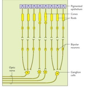

- Outer pigmented layer
- A layer of rods and cones (photoreceptor cells)
- External limiting membrane
- Outer nuclear layer (including cell bodies of rods and cones)
- Outer plexiform layer
- Inner nuclear layer (Cell bodies of bipolar neurons)
- Inner plexiform layer
- Ganglion cell layer
- Nerve fiber layer
- Internal limiting membrane
Vitreous body
It is a clear, transparent jelly-like substance that fills the eyeball behind the lens (99% water, with some salts and meshwork of collagenous fibers, mucopolysaccharide, hyaluronic acid) The functions are; supports the posterior surface of the lens and helps maintains intraocular pressure in the eye.
Lens
Unusual biological structure. Thick, transparent, biconvex disc and circular in outline. Held in place by its attachment with the ciliary process of the ciliary body with the help of the suspensory ligament. The external Features: Anterior and posterior surfaces, Anterior and posterior poles, and A circumference-the equator.
Clinical aspects of eye
- Glaucoma
- Presbyopia (short vision)
- cataract
- Horner Syndrome
[embeddoc url=”https://notesmed.com/wp-content/uploads/2021/05/An-Eye.pdf” download=”all” cache=”off”]