What is the elbow joint?
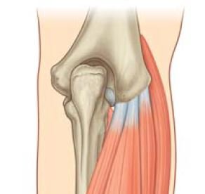
Elbow joint is a hinge type of synovial joint and present between the lower end of the humerus bone and upper ends of the radius and ulnar bone. It literally includes two articulations:
- Humero-ulnar articulation:
- It is present between the trochlea of the humerus bone and the trochlear notch of the ulna bone.
- Humero-radial articulation:
- It is present between the capitulum of the humerus bone and the head of the radius bone.
On the surface, the elbow joint which a line of the elbow situated about 2 cm below the line joining the two epicondyles of the humerus bone. The complexity of this elbow joint is furthermore increased by its continuity with the superior (upper) radio-ulnar joint. There are three articulations present in the elbow region, viz.
- Humero -ulnar
- Humero-radial, and
- Superior (proximal) radio-ulnar.
The elbow joint is also known as cubital articulations.

Articular surfaces
- Upper articular surface: It formed by the capitulum and the trochlea of the lower extremity of the humerus bone.
- Lower articular surface: It formed by the superior surface of the head of the radius bone and the trochlear notch of the ulna.
- Capitulum:
- It is a rounded hemispherical eminence that possesses a smooth articular surface area only on its anterior and inferior aspects.
- Trochlea:
- It is medial to the capitulum in the bone & bears a resemblance to a pulley. The medial flange of the trochlea which is projects to an inferior (lower) level than its lateral flange.
- The trochlear notch of the ulna:
- It is composed of the upper surface of the coronoid process and the anterior surface of the olecranon process the ulna.
- The upper end of radius:
- It is circular in outline present and slightly depressed area in the center.
Ligaments
- Capsular ligament (Joint capsule).
- Medial ligament (Ulnar collateral ligament).
- Lateral ligament (Radial collateral ligament).
Relations
Anterior:
- Brachialis muscle.
- Median nerve.
- Brachial artery.
- Tendon of biceps brachii.
Posterior:
- Tendon of the triceps muscle
- Anconeus muscle
Medially:
- Flexor carpi ulnaris
- Ulnar nerve (posteromedially)
- Common flexor origin of the muscles of the forearm in anteromedially.
Laterally (posterolateral):
- Supinator
- Common extensor origin of muscles of the forearm
- Extensor carpi radialis brevis muscle
Bursae related to the elbow joint
- Four important bursae are present in this joint, they are following;
-
- The subtendinous olecranon bursa:
- It is a small bursa present between the triceps tendon and the upper surface of the olecranon process of the ulnar bone.
- The subcutaneous olecranon bursa:
- It is a large bursa present between the skin area and subcutaneous triangular area on the posterior surface of the olecranon.
- Bicipitoradial bursa:
- It is a small bursa separating the biceps tendon from the smooth or plane anterior part of the radial tuberosity.
- A small bursa that separating or dividing the biceps tendon from the oblique cord.
- The subtendinous olecranon bursa:
Stability of the elbow joint
- The pulley-shaped trochlea of the humerus fits correctly into the jaw-like trochlear notch of the ulnar bone.
- Strong ulnar ligament and radial collateral ligament
Arterial supply
It is supplied by arterial anastomosis around the elbow joint is formed by the branches of the brachial artery, radial, & ulnar arteries.
Nerve supply
By articular branches from:
- Radial nerve (through its branch to anconeus),
- Musculocutaneous nerve (through its branch to brachialis),
- The ulnar nerve, and
- Median nerve.
Movements of the elbow joint
- Flexion: (muscle)
-
- Brachialis
- Biceps brachii
- Brachioradialis
- Extension: (muscle)
- Triceps
- Anconeus
Applied anatomy
- Elbow effusion.
- Dislocation of the elbow joint.
- Nursemaid’s elbow/pulled elbow (subluxation of the head of the radius bone).
- Tennis elbow (lateral epicondylitis).
- Golfer’s elbow (medial epicondylitis).
- Student’s elbow (Miner’s elbow).
- Nerve entrapments (compressions) around the elbow joint.
[embeddoc url=”https://notesmed.com/wp-content/uploads/2020/08/ELBOW-JOINT.pdf” download=”all”]