What is the ear?
The ear is the most important organ of hearing and plays an important role in maintaining the balance (equilibrium) of the body.
Parts
The ear is divided into three parts, are following;
- External ear
- Auricle/pinna
- External auditory meatus
- Middle ear/tympanic cavity
- Internal ear

External ear
The external ear consists of the auricle and external auditory meatus at the medial end of which lies the tympanic membrane or eardrum, separating the external ear from the middle ear.
Auricle/pinna
The auricle is a trumpet-like undulating projection present on the side of the head. The whole pinna except its lobule is made up of a single piece of scrumpled yellow elastic cartilage surrounded with skin.
The lobule of the pinna is made of fibrofatty tissue and surrounded by skin. The auricular cartilage is continual with the cartilage of the external auditory meatus.
Features of auricle
The lateral surface of the auricle shows the following elevations and depressions, they are concha, helix, antihelix, tragus, antitragus, cymba conchae, and lobules.
The medial/ cranial surface of the pinna has the following features such as eminentia concha and ementia triangularis. No cartilage present between tragus and crus and the gap present between the two called Incisura terminalis.
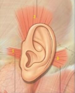
Muscle of the auricle
The muscles of the auricle are divided into two sets: extrinsic and intrinsic but they are rudimentary in humans being. The extrinsic muscles pass from the scalp or skull to the auricle and they are; anterior, posterior, and superior auricularis muscles.
The intrinsic muscles of the auricle are small muscular slips, which pass between the cartilaginous parts of the auricle. The pinna collects and directs the sound waves to transfer into the external auditory meatus.
External auditory meatus
The acoustic meatus extends from the bottom of the concha to the tympanic membrane and which is not a straight tube but it has a typical ‘S’ shape course and measures a length of about 24 mm along its posterior wall.
The external auditory meatus is divided into two parts such as lateral 1/3rd cartilaginous continuous with the cartilage of the auricle and medial 2/3rd part is bony. The skin covering the cartilaginous part is thick and contains hair and ceruminous (pilosebaceous) glands, which secrete ear wax but the bony part doesn’t contain it.
How to observed external auditory meatus?
The observation of the external acoustic meatus and tympanic membrane can be improved by pulling the ear superiorly, posteriorly, and slightly laterally.
Tympanic Membrane (eardrum)
The eardrum is a thin, semitransparent partition(division) between the external auditory canal and the middle ear of the body. It is an oval, measuring 9–10 mm in length, and 8–9 mm in width.
The eardrum/tympanic membrane obliquely placed making an angle of about 55° with the floor of the external acoustic meatus. It consists of a connective tissue core bounded with skin on the outside and mucous membrane on the inside.
Structure of the tympanic membrane
The tympanic membrane contains three layers; from lateral to medial these are as follows:
- Outer cuticular layer of stratified squamous epithelium:
- Continuous with the skin bounding the external auditory, meatus.
- Middle fibrous layer, which surrounds the handle of the malleus.
- Contains outer radiating and inner circular fibers.
- The Innermucosal layer is lined by low columnar epithelium
- Continuous with the mucous surrounding of the middle, ear.
Parts of the tympanic membrane
- Pars tensa: It is most of the tympanic membrane and periphery is thickened to form a fibrocartilaginous rim called annulus tympanicus, which fits into the tympanic sulcus.
- Pars flaccida (Shrapnell’s membrane): a small triangular area above the lateral process of malleus between anterior and posterior malleolar folds (now called malleal folds).
Surfaces of the tympanic membrane
- Lateral surface: concave towards the meatus and directed downwards, forwards, and laterally.
- Medial surface: convex and bulges towards the middle ear. The umbo is the point of maximum convexity.
Middle ear (tympanic cavity)
The middle ear is a narrow slit-like air-filled cavity within the petrous part of the temporal bone. It is sandwiched between the external and internal ears. The shape is a cube, compressed from side to side. In the coronal section, it resembles a biconcave disc, like a red blood cell.
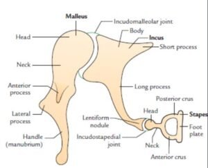

Parts of the middle ear are the epitympanic recess and tympanic cavity proper. It consists of three bones called ear ossicles: malleus, incus, and stapes. And it also contains two muscles (tensor tympani and stapedius), two nerves (chorda tympani and tympanic plexus), vessels supplying and draining the middle ear and ligaments of the ear ossicles.
Communications of the middle ear
Anteriorly communicates with the nasopharynx through the eustachian tube. Posteriorly communicates with mastoid antrum and mastoid air cells through aditus to antrum called aditus ad antrum.
Walls (boundaries) of the middle ear
- Roof or tegmental wall
- Floor or the jugular wall
- Anterior or the carotid wall
- Posterior or the mastoid wall
- Lateral or the membranous wall
- Medial or the labyrinthine wall
Features of roof or tegmental wall
It is formed by a thin plate of bone called tegmen tympani so-called tegmental wall. It partitions the tympanic cavity from the middle cranial fossa. The tegmen tympani also extend posteriorly to form the roof of the aditus ad antrum.
Features of floor or jugular wall
It is formed by a thin plate of the bone and which separates the tympanic cavity from the jugular bulb so-called jugular wall.
Features of the anterior wall of carotid wall
It is formed by a thin plate of bone. The lower part separates the cavity from the internal carotid artery and the upper part of the anterior wall presents two openings or canals, the upper one for the tensor tympani muscle & the lower one for the external auditory tube the ear.
Features of the posterior wall or mastoid wall
The posterior wall contains the following structures such as aditus ad antrum, fossa incudis, pyramid, vertical part of the facial canal, and posterior canaliculus for chorda tympani.
Features of the medial or the labyrinthine wall
The medial or the labyrinthine wall contains the following structures as
- Promontory
- Oval window (fenestra vestibuli)
- Round window (fenestra cochleae)
- Sinus tympani
- The prominence of the oblique part of the facial canal
The prominence of the lateral semicircular canal of the internal ear.
Features of the lateral or the membranous wall
It is formed by the tympanic membrane, which separates the tympanic cavity from the external auditory meatus. The chorda tympani nerve, which is a branch of the facial nerve passes over the tympanic membrane lying lateral to the long process of the incus and medial to the handle of the malleus.
Internal Ear
The internal ear consists of a closed system of fluid-filled intercommunicating membranous sacs and ducts called membranous labyrinth (endolymph). The membranous labyrinth lies within the complex intercommunicating between bony cavities and canals (bony labyrinth) in the petrous part of the temporal bone.
The space present between the membranous and bony labyrinth is filled with fluid called perilymph. The components of the internal ear viz. membranous labyrinth and bony labyrinth.
Membranous labyrinth
The membranous labyrinth consists of the following parts such as cochlear duct, saccule, utricle, and semicircular ducts (three). The cochlear duct lies within the bony cochlea, the saccule, and the utricle lies within the bony vestibule, and three semicircular ducts lie within the three bony semicircular canals.
Cochlear Duct (Scala Media)
The cochlear duct lies in the middle part of the cochlear canal and contains the spiral organ of the Corti (sensory receptor for hearing). In the cross-section of the cochlear canal- the cochlear duct is triangular in shape. The boundaries are following
- Base: It is formed by the osseous spiral lamina which is a medial and the basilar membrane which is lateral.
- Roof: Formed by the vestibular membrane (Reissner’s membrane).
- Laterally: by the outer wall of the cochlear canal.
Spiral Organ of Corti
It is a peripheral organ of hearing in the cochlear duct. It is situated on the basilar membrane. The components are tunnel of Corti, Hair cells, Supporting cells(Deiter’s and Hansen’s Cells), and membrana tectoria.
Bony labyrinth
It consists of a series of intercommunicating bony cavities and canals within the petrous part of the temporal bone. The bony labyrinth presents three parts from before backward these are cochlea, the vestibule, and the semicircular canal (3).
Modiolus is an axial bony stem around which the cochlear canal spirals and it is like an elongated cone.
The Cochlear canal arranged spirally around the modiolus and makes two and three-fourth turns. Its basal turn bulges into the tympanic cavity as the promontory and its cavity as the promontory.
Spiral lamina is a triangular area thus enclosed by the vestibular and basilar membranes, and the outer wall of the cochlear canal from the cochlear duct (scala media). Scala vestibuli and scala tympani communicate with one another at the apex of the cochlea by a small opening called helicotrema.
At the basal turn of the cochlea, the scala vestibule communicates with the anterior wall of the vestibule. And close to the basal turn of the cochlea, the scala tympani presents two features fenestra cochleae and the beginning of the aqueduct of the cochlea.
The vestibule is a bony cavity between the cochlea and semicircular canal.
- Anterior wall-opening of the cochlear canal
- Posterior wall– 5 openings of semicircular canal
- Lateral wall– Fenestra vestibuli
- Medial wall-spherical recess and elliptical recess and two recesses are separated by the vestibular crest which splits inferiorly to enclose the cochlear recess.
Semicircular canals
The semicircular canals are three canals (anterior (superior), posterior, and lateral) lie in three planes at a right angle to each other. Open in the vestibule by 5 openings and dilated one end is called the ampulla.
Anterior semicircular canal-right angle to the long axis of the petrous part of the temporal bone and posterior semicircular canal- parallel long axis of the petrous part of the temporal bone and lateral semicircular canal lies in the horizontal plane.
Applied anatomy
- Infection of the middle ear (otitis media)
- Acute mastoiditis and mastoid abscess
- Meningitis and temporal lobe abscess
- Lower motor neuron type of facial palsy
- Transverse and sigmoid sinus thrombosis
- Labyrinthitis
- Cerebellar abscess
- Hyperacusis
- Paralysis of stapedius muscle that results in hyperacusis (an abnormally increased power of hearing) where even whisper appears as noise.
- Otosclerosis
- Abnormal ossification of the annular ligament, which anchors the footplate of stapes to the oval window is called otosclerosis and impedes the movements of stapes and causes deafness. It is the most common cause of conductive deafness in adults.
- Ramsay Hunt syndrome (Involvement of pinna in herpes zoster of geniculate ganglion)