What is the duodenum?
The term “duodenum” is obtained from the Latin corruption of the Greek word “do-deka-daktulos” meaning 12 fingers.
The duodenum is the shortest, broadest(widest), and most fixed part of the small intestine. It extend from the pylorus to duodenojejunal flexure. The length of the duodenum is about 10 inches (25 cm) long.
It starts at the pylorus of the stomach which lies on the transpyloric plane around 2.5 cm to the right of the median plane & ends at the duodenojejunal junction which lies around 2.5 cm to the left of the median plane and a little beneath the transpyloric plane.
The main significance of the duodenum is the digestion of the food. It is C- shaped structure, near the head of the pancreas in the human.
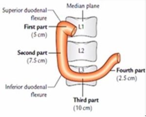
It located in the abdominal cavity above the level of the umbilicus; opposite the first (L1), second (L2), and third (L3) lumbar vertebrae.

Parts of the duodenum
The duodenum divided into four parts, they are the following;
- Superior (first) part, 5 cm (2 inches) long.
- Descending (second) part, 7.5 cm (3 inches) long.
- Horizontal (third) part, 10 cm (4 inches) long.
- Ascending (fourth) part, 2.5 cm (1 inch) long.
Superior (first) part:
Its expanse (extent) from the pylorus of the stomach to superior duodenal flexure. The main characteristics of this part are the following;
- It develops from the foregut.
- It is only partly retroperitoneal part.
- It is freely mobile (motile) and distensible.
- In the mucus membrane, there are no circular folds of its initial 2.5 cm-seen as the duodenal cap in barium meal on radiographs.
- It is the area for duodenal ulcers.
- It is supplied by the branches of the coeliac trunk/artery.
Relations of superior (first) part of the duodenum:


Relations of superior (first) part of the duodenum
- Anteriorly: Quadrate lobe of the liver and gallbladder.
- Posteriorly: Related to the portal vein, gastroduodenal artery, and common bile duct (CBD).
- Superiorly: Epiploic foramen being divided from it by means of the portal vein and bile duct.
- Inferiorly: Head & neck of the pancreas.
Descending (second) part
It extends from superior duodenal flexure(L1) to inferior duodenal flexure (L3). The main features of the second part of the duodenum are;
- Its upper half develops from the foregut and the lower half from the midgut.
- It lies behind the transverse mesocolon.
- It receives the bile duct, the chief and accessory pancreatic ducts.
- It is the only part of the intestine supplied by double rows of the vasa recta, arising from anterior and posterior pancreaticoduodenal arterial arcades.
Relations of descending (second) part of the duodenum
Relations of descending (second) part of the duodenum
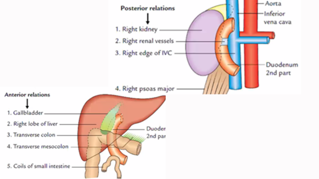
- Anteriorly: It is related to the Gallbladder and liver (right lobe), transverse colon and mesocolon (commencement), and coils of the small intestine.
- Posteriorly: Right kidney and right renal vessels, the right edge of the inferior vena cava (IVC), and right psoas major muscle.
- Medially: Head of the pancreas.
- Laterally: From beneath upward; ascending colon, right colic flexure, & right lobe of the liver.
Interior of the duodenum(second part)
- Circular folds
- Major duodenal papilla: Clearly defined conical projection on the posteromedial wall of the duodenum and located 8–10 cm distal to the pylorus.
- Minor duodenal papilla: A small conical projection pinpoint 2 cm proximal (and ventral) to the major duodenal papilla.
- Arch of plica semicircularis: It forms an arch above the major duodenal papilla like a hood (cf. monk’s hood).
- Plica longitudinalis: It is a vertical tortuous fold of the mucous membrane extending downward from the major duodenal papilla.
Horizontal (third) part
It extends from the inferior duodenal flexure to the front of the aorta (L3). The relations of the horizontal (third) part of the duodenum is;

- Anteriorly: Root of the mesentery, superior mesenteric vessels (SMV), & coils of the jejunum.
- Posteriorly: Right psoas major muscle, right ureter, Inferior vena cava (IVC), abdominal aorta, and right gonadal vessels.
- Superiorly: The uncinate process of the head of the pancreas.
- Inferiorly: Coils of the jejunum.
Ascending (fourth) part
It extends from the front of the aorta to duodenojejunal flexure (L2). The relations of ascending (fourth) part of the duodenum is:

- Anteriorly: Transverse colon and mesocolon.
- Posteriorly: Left psoas major muscle, left sympathetic chain, left gonadal vessels, and inferior mesenteric vein (IVC).
- Superiorly: Body of the pancreas.
- On to the left: Left side kidney and left ureter.
- On to the right: Upper part of the root of the mesentery.
Suspensory muscle of the duodenum (LIGAMENT OF TREITZ)
It is a fibromuscular band, which suspends the duodenojejunal flexure from the right crus of the diaphragm.
Its upper end is attached to the right crus of the diaphragm and the lower end attached to the posterior surface of the duodenojejunal flexure.
This band contains striated muscle fibers in the upper part, elastic fibers in the middle part, and non-striated muscle fibers in the lower part.
Duodenal recesses (fossae)
The duodenojejunal junction, small pocket-like pouches of peritoneum called duodenal recesses. They are;
- Superior duodenal recess.
- Inferior duodenal recess.
- Paraduodenal recess.
- Retroduodenal recess.
Arterial supply
- Superior pancreaticoduodenal artery.
- Inferior pancreaticoduodenal artery.
- Supraduodenal artery of “Wilkie”.
- Retroduodenal branches of the gastroduodenal artery.
- Leash of branches of the hepatic artery.
- Branches from the right gastroepiploic artery.
- An artery from the first jejunal branch of the superior mesenteric artery.
Venous drainage:
- Splenic vein.
- Superior mesenteric vein
- Portal veins.
Lymphatic drainage:
- Pancreaticoduodenal nodes.
- Coeliac and a mesenteric group of the lymph nodes.
Nerve supply:
- Sympathetic nerves: T6–T9 segments of the spinal cord.
- Parasympathetic nerves: from both the vagi through coeliac and superior mesenteric plexuses in the duodenum.
Microscopy of the duodenum:
The duodenum has four layers, they are:

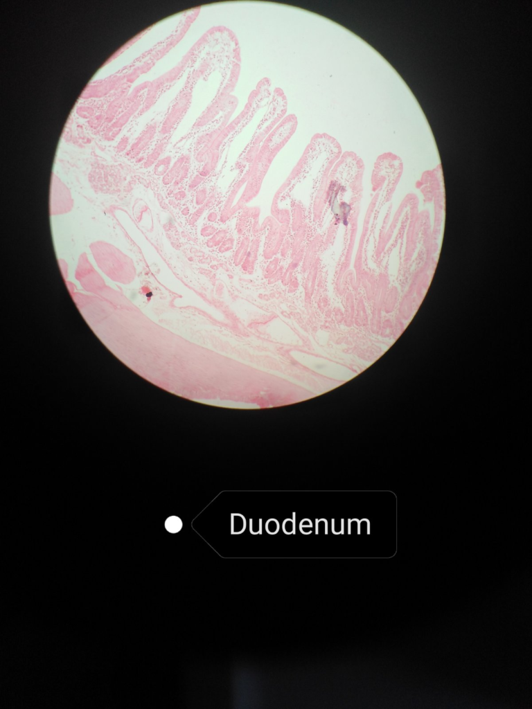
- Mucosa:
- It is lined by simple columnar epithelium, lamina propria, and muscular mucosae with connective tissue cells, lymphatic cells, plasma cells, smooth muscle cells, etc.
- The villi are leaf-like, tall, and numerous with few goblet cells in the epithelium.
- Submucosa:
- It consists of the Brunner’s gland.
- Muscularis Externa:
- It consists of an inner circular layer and outer longitudinal smooth muscle layers.
- Serosa (visceral peritoneum):
- It contains connective tissue cells, blood vessels, and adipose cells. The outermost layer of the first part of the duodenum.
[embeddoc url=”https://notesmed.com/wp-content/uploads/2021/02/DUODENUM.pdf” download=”logged”]