What is the diaphragm?
The thoracic outlet is closed by a large dome-shaped musculotendinous structure which is known as the diaphragm. The diaphragm is the primary muscle that involved in the respiration process.
Origin: three parts, viz.
- Sternal part: It consists of 2 fleshy slips, which are arising from the posterior surface of the xiphoid process.
- Costal part: On each side, it consists of 6 fleshy slips, which are arising from the inner surface of the lower six ribs near their costal cartilages of the body.
- Vertebral part: These parts are arising by means of the following structures;
- Right crura & left crura of the diaphragm with five arcuate ligaments.
Crura:
| Right crus | Left crus |
| Vertical fleshy bundles mass | Vertical fleshy bundles mass |
| Arising from the right side of anterior aspects of the upper three lumbar (L1-L3) vertebrae. | Arising from the left side of anterior aspects of the upper two lumbar(L1 and L2) vertebrae. |
| Intervening intervertebral discs in the body. | Intervening intervertebral discs in the body |
Arcuate ligaments:
- Median arcuate ligament: It is an arched fibrous band which is stretching between the upper extremities (ends) of two crura.
- Medial arcuate ligament: It is the thickened superior margin of the psoas sheath and that extends from the side of the body of the second lumbar (L2) vertebra to the tip of the transverse process of the first lumbar (L1) vertebra.
- Lateral arcuate ligament: It is the thickened superior margin of fascia that covering the anterior surface of the quadratus lumborum muscle and extends from the tip of the transverse process of the first lumbar (L1) vertebra to the 12th rib in the body.

Insertion:
From circumferential origin (vide supra), the muscle fibers intersect towards the central tendon & insert into its margins.
The characteristics of the central tendon are the following below;
- It is trifoliate in shape structure, having (a) anterior (central) leaflet structures, and (b and c) two tongue-shaped posterior leaflet structures. It bears a resemblance to an equilateral triangle. The right posterior leaflet is a short & stout structure, whereas the left posterior leaflet is a thin & long structure in the body.
- It is inseparably merged with fibrous pericardium.
- It is situated nearer to the sternum than to the vertebral column in the body.
Surfaces of diaphragm
- Superior surface:
- It projects on either side as a dome-shaped or cupola into the thoracic cavity.
- The depressed area present between the two domes is called a central tendon.
- The superior(upper) surface is surrounded by endothoracic fascia.
- Inferior surface:
- It lined by the diaphragmatic fascia and parietal peritoneum in the body.
Relations of the diaphragm
Superior surface related to:
- The bases of the right pleura and left pleura on the sides.
- The fibrous pericardium is in the middle region.
Inferior surface related to:
On the right side, it is related to the following structures:
- The right lobe of the liver.
- Right kidney.
- Right suprarenal gland.
On the left side, it is related to the following structures:
- Left lobe of the liver.
- Fundus of the stomach.
- Spleen.
- Left kidney.
- Left suprarenal gland.
Openings of the Diaphragm
The openings of the diaphragm are classified into two types, they are following below:
- Major openings
- Minor openings.
Major openings
There are three major openings are present, viz.
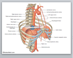
- Vena caval opening.
- Esophageal opening.
- Aortic opening.
| Opening | Location | Shape | Vertebral level |
| Vena caval opening | In the central tendon, it is slightly to the right of the median plane present between the central and right posterior leaflets. | Quadrangular or square | T8 (body) |
| Esophageal opening | Slightly to the left of the median plane (The fibers of the right crus splits around the opening and act like a pinchcock). | Oval or elliptical | T10 (body) |
| Aortic opening | In the midline beyond the median arcuate ligament in the body. | Circular or round-shaped | T12 (lower border of the body) |
| Opening | Structures passing through |
| Vena caval opening | • Inferior vena cava. • Right phrenic nerve |
| Esophageal opening | • Esophagus. • Right and left vagal trunks. • Esophageal branches of the left gastric artery. |
| Aortic opening | From right to left these are the following: – Azygos vein. – Thoracic duct.– Aorta. |
Minor Openings
- Superior epigastric vessels which are pass through the gap (space of Larry) present between the muscular slips that arising from the xiphoid process and the 7th costal cartilage.
- The musculophrenic artery which it passes through the gap present between the slips of origin from the 7th to the 8th ribs in the diaphragm.
- Lower 5 intercostal nerves and vessels (i.e., 7th to 11th) that pass through gaps present between the adjoining costal slips in the diaphragm.
- Subcostal nerves and vessels which are pass deep to the lateral arcuate ligament.
- The sympathetic chain which is passes deep to the medial arcuate ligament.
- Greater, lesser, and least splanchnic nerves which are pass by penetrating the crus of the diaphragm on the corresponding side.
- hemiazygos vein penetrates the left crus of the diaphragm.
Actions of Diaphragm
- A muscle of inspiration: main/principal muscle involved in the respiration process. When it contracts, it descends downwards and increases the vertical diameter of the thoracic cavity.
- A muscle of abdominal staining: (voluntary expulsive efforts, e.g., micturition, defecation, vomiting, parturition, etc.).
- A muscle of weight lifting: taking a deep breath & closing the glottis in the body.
- Thoraco-muscular pump.
- Sphincter of the esophagus.
Development of diaphragm

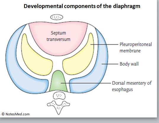
- Septum transversum (ventrally).
- Pleuroperitoneal membranes at the sides.
- The dorsal mesentery of the esophagus (dorsally).
- Body wall (peripherally).
- The central tendon of the diaphragm is developed from the septum transversum.
- Domes of the diaphragm are developed from the pleuroperitoneal membrane.
- Part of the diaphragm around the esophagus is developed from the dorsal mesentery of the esophagus of the body.
- The peripheral part of the diaphragm is developed from the body wall.
Applied anatomy
- Diaphragmatic paralysis (paralysis of the diaphragm):
- It is unilateral damage of the phrenic nerve in the body that leads to unilateral diaphragmatic paralysis.
- Hiccups:
- It occurs due to involuntary spasmodic contractions of your diaphragm which is along with the closure of the glottis.
- Hiccups generally occur after eating foods or drinking fluids as a result of gastric irritation.
- Pathological causes include following diaphragmatic irritation, phrenic nerve irritation, hysteria, and uremia.
- Diaphragmatic hernias (Congenital and acquired).
[embeddoc url=”https://notesmed.com/wp-content/uploads/2020/08/Thorax.pdf” download=”all” cache=”off”]