What is axilla ?
The axilla or armpit is a fat-filled pyramid-shaped space, between the upper part of the arm and the side of the chest wall. It contains the brachial plexus, axillary vessels, and lymph nodes.
Boundaries
The axilla resembles a truncated four-sided pyramid and presents an apex, a base, and four walls (anterior, posterior, medial, and lateral).

Apex
It is also called the cervico-axillary canal, triangular in shape. It is a passageway between the neck and axilla.
It is directed upward and medially towards the root of the neck. Bounded in front by the clavicle, behind by the superior border of the scapula, and medially by the outer border of the first rib.
Axillary vessels and the cord of the brachial plexus passes through the apex.
Base or Floor
It is at the lower end of the axilla and directed below and present a concavity which is bounded-
In front by anterior Axillary fold, formed by the lower border of the pectoralis major muscles.
Behind by the posterior axillary fold formed by the tendon of latissimus dorsi and teres major muscles.
Medially by a lateral aspect of the chest wall.
In addition, it is formed by skin, superficial fascia, and axillary fascia.
Anterior wall
It is formed by the pectoralis major and pectoralis minor, subclavius. The anterior axillary fold is the inferior most part of the anterior formed by the pectoralis major.
Posterior wall
It is formed by the Subscapularis muscle above, Teres major, and latissimus dorsi below. The posterior axillary fold is the inferior most part of the posterior wall.
Medial wall
It is broad and formed by the upper four or five ribs and their intercostal muscles and the upper part of the serratus anterior muscle.
Lateral wall
It is narrow and both anterior and posterior walls of the axilla converge on it. It is formed by –Intertubercular sulcus of the shaft of the humerus which contains a long tendon of biceps brachii and tendons of coracobrachialis and short head of biceps brachii.
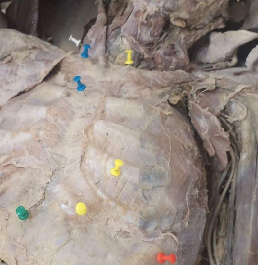
Contents of the axilla
- Axillary artery and its branches.
- Axillary vein and it tributaries
- Cords of brachial plexus and their branches
- Long thoracic nerve and intercostobrachial nerve
- Axillary lymph nodes
- Axillary fat (fibrofatty tissue)
- The axillary tail of the breast (Spence).
Axillary lymph nodes:
- Anterior or pectoral group: lie along the lateral thoracic vein at the lower border of the pectoralis minor.
- Posterior or subscapular group: lie on the posterior axillary fold along the subscapular vein.
- Lateral group: lie along the upper part of the humerus in relation to the axillary vein.
- Central group: situated in the upper part of the axilla.
- Apical or infraclavicular group: situated deep to the clavipectoral fascia at the apex of the axilla along the axillary vein.
Applied anatomy
- Enlargement of Axillary Lymph Nodes
An infection in the upper limb can cause the axillary nodes to enlarge and become tender and inflamed, a condition called lymphangitis
- Surgical approach to axilla
The axilla is approached surgically through the skin of the floor of the axilla.
- Brachial Plexus Injuries (Mainly cords and their branches)
- Axillary abscess