
What is acute appendicitis?
Acute appendicitis is an acute inflammation of the vermiform appendix in the human body. It is the most common cause of abdominal emergency in both adults as well as children.
Etiopathogenesis
The etiology of appendicitis is multifactorial and may involve various factors such as obstruction, ischemia, infections, and hereditary factors.
In children, Lymphoid hyperplasia (60% of cases) often secondary to viral infection. Virus-like Adenovirus, measles virus infection, or immunization.
In adults, Fecalith obstructs the proximal lumen so increased intraluminal pressure causes mucosal injury and bacterial invasion in an appendix and other causes are gallstones, tumors, a mass of worms (Oxyuriasis vermicularis), etc.
It is initiated by progressive increases in intraluminal pressure that compromise venous outflow. Stasis of luminal contents – favors bacterial proliferation, triggers ischemia and inflammatory responses, and finally results in tissue edema and neutrophilic infiltration in lumen, muscular wall as well as peri-appendiceal soft tissues.
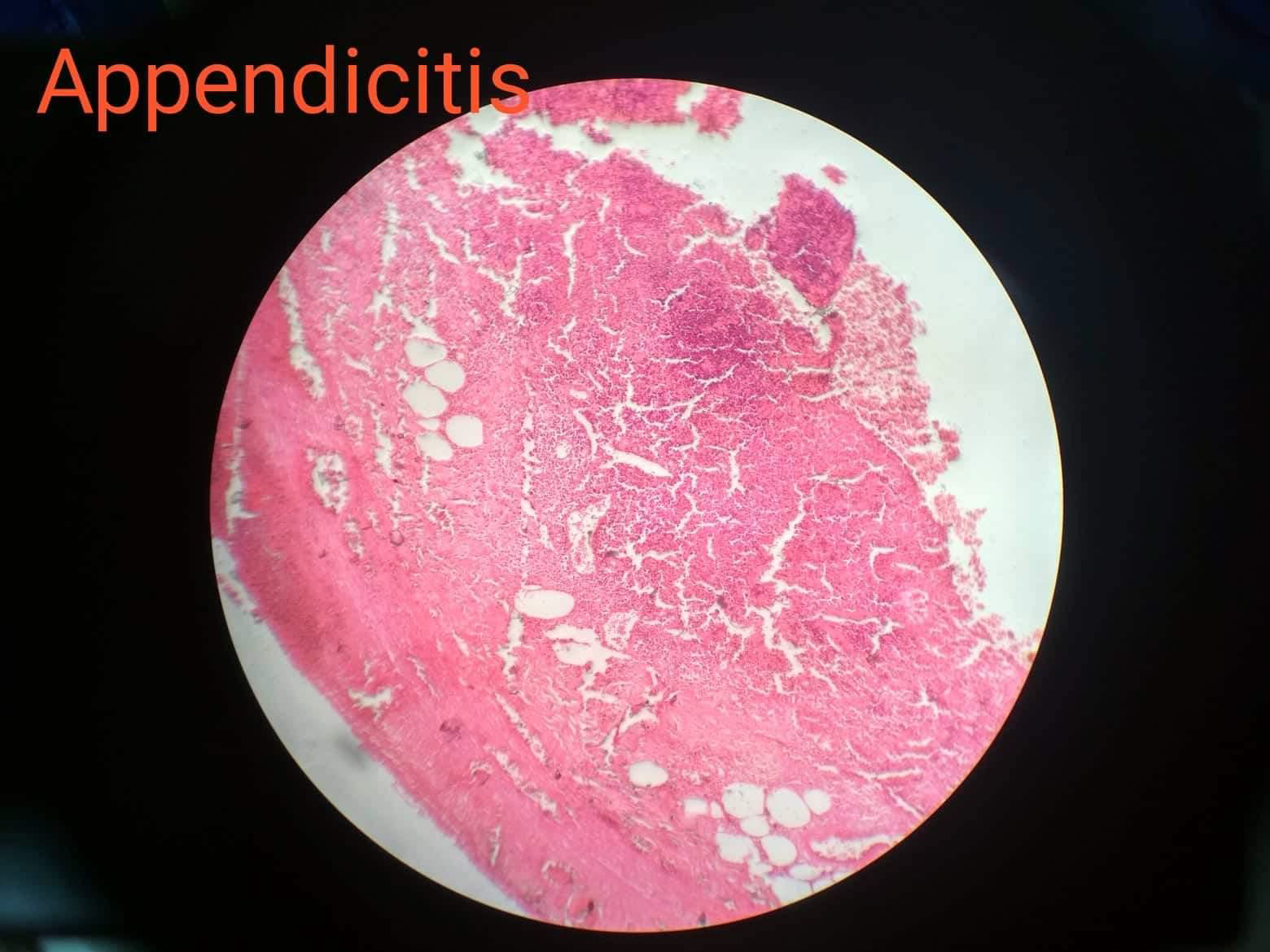
Morphology
Gross
- Normal glistening serosa becomes dull, granular, and erythematous
- Fibrinous or purulent coating of serosa
- Engorgement of subserosal vessels
- Mucosal ulceration
- Gangrenous: greenish black covered with exudate
- Obstruction of the lumen by Fecolith or some other agent in nearly 25-33% of cases.

Microscopy
Three stages are present, they are;
- Acute(early) Appendicitis
- Acute Suppurative Appendicitis
- Acute Gangrenous Appendicitis

Early lesions Appendicitis: Modest perivascular neutrophilic infiltrate within all layers of a wall with Congested subserosal vessels.
Later stages (Acute Suppurative Appendicitis): Prominent neutrophilic exudate and abscess formation, Ulceration, Suppurative necrosis in the mucosa can be seen.
Acute Gangrenous Appendicitis: Mucosal ulceration and gangrenous necrosis throughout the wall.
The diagnosis of appendicitis must require neutrophilic infiltration in muscularis propria. Sometimes a prominent histiocytic component with clusters of xanthoma-type cells can be seen in microscopy is called xanthogranulomatous appendicitis.
Complications
- Perforation of wall
- Peritonitis
- Fibrosis
- Hemorrhage
- Appendicular abscess
- Fistula tract
- Septicaemia etc.
McBurney’s sign: It is a physical examination of deep tenderness located two-thirds of the distance from the umbilicus to the right anterior superior iliac spine (McBurney’s point).
[embeddoc url=”https://notesmed.com/wp-content/uploads/2021/05/Appendicitis.pdf” download=”all” text=”Complete pdf file-Download”]
